More info
Overview
Long Name | Antibody Type | Antibody Isotype | Host | Species Reactivity | Validated Applications | Purification |
| signal transducer and activator of transcription 1, 91kDa | Polyclonal | IgG | Rabbit | Human, Mouse, Rat | WB | Immunogen affinity purified. |
Immunogen | ||||||
| A synthetic peptide corresponding to a sequence in the middle region of human STAT1(364-378aa FDKDVNERNTVKGFR), different from the related mouse sequence by one amino acid. | ||||||
Properties
Form | Lyophilized |
Size | 100 µg/vial |
Contents | Antibody is lyophilized with 5 mg BSA, 0.9 mg NaCl, 0.2 mg Na2HPO4, 0.05 mg Thimerosal and 0.05 mg NaN3. *carrier free antibody available upon request. |
Concentration | Reconstitute with 0.2 mL sterile dH2O (500 µg/ml final concentration). |
Storage | At -20 °C for 12 months, as supplied. Store reconstituted antibody at 2-8 °C for one month. For long-term storage, aliquot and store at -20 °C. Avoid repeated freezing and thawing. |
Additional Information Regarding the Antigen
Gene | STAT1 |
Protein | Signal transducer and activator of transcription 1 |
Uniprot ID | P42224 |
Function | Signal transducer and transcription activator that mediates cellular responses to interferons (IFNs), cytokine KITLG/SCF and other cytokines and other growth factors. Following type I IFN (IFN-alpha and IFN-beta) binding to cell surface receptors, signaling via protein kinases leads to activation of Jak kinases (TYK2 and JAK1) and to tyrosine phosphorylation of STAT1 and STAT2. The phosphorylated STATs dimerize and associate with ISGF3G/IRF-9 to form a complex termed ISGF3 transcription factor, that enters the nucleus. ISGF3 binds to the IFN stimulated response element (ISRE) to activate the transcription of IFN- stimulated genes (ISG), which drive the cell in an antiviral state. In response to type II IFN (IFN-gamma), STAT1 is tyrosine- and serine-phosphorylated. It then forms a homodimer termed IFN- gamma-activated factor (GAF), migrates into the nucleus and binds to the IFN gamma activated sequence (GAS) to drive the expression of the target genes, inducing a cellular antiviral state. Becomes activated in response to KITLG/SCF and KIT signaling. May mediate cellular responses to activated FGFR1, FGFR2, FGFR3 and FGFR4. |
Tissue Specificity | |
Sub-cellular localization | Cytoplasm. |
Sequence Similarities | Belongs to the transcription factor STAT family. |
Aliases | Signal transducer and activator of transcription 1 91kD antibody|DKFZp686B04100 antibody|ISGF 3 antibody|ISGF-3 antibody|OTTHUMP00000163552 antibody|OTTHUMP00000165046 antibody|OTTHUMP00000165047 antibody|OTTHUMP00000205845 antibody|Signal transducer and activator of transcription 1 91kDa antibody|Signal transducer and activator of transcription 1 alpha/beta antibody|Signal transducer and activator of transcription 1 antibody|Signal transducer and activator of transcription 1, 91kD antibody|Signal transducer and activator of transcription 1-alpha/beta antibody|Signal Transductor and Activator of Transcription 1 antibody|STAT 1 antibody|STAT 91 antibody|Stat1 antibody|STAT1_HUMAN antibody|STAT91 antibody|Transcription factor ISGF 3 components p91 p84 antibody|Transcription factor ISGF-3 components p91/p84 antibody |
Application Details
| Application | Concentration* | Species | Validated Using** |
| Western blot | 0.1-0.5μg/ml | Human, Mouse, Rat | AssaySolutio's ECL kit |
AssaySolution recommends Rabbit Chemiluminescent WB Detection Kit (AKIT001B) for Western blot. *Blocking peptide can be purchased at $65. Contact us for more information
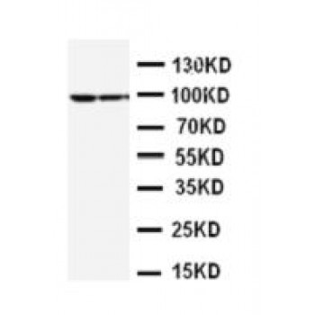
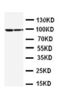
Anti-STAT1 antibody, ASA-B1789, Western blotting
Lane 1: MCF-7 Cell Lysate
Lane 2: HELA Cell Lysate
Lane 1: MCF-7 Cell Lysate
Lane 2: HELA Cell Lysate
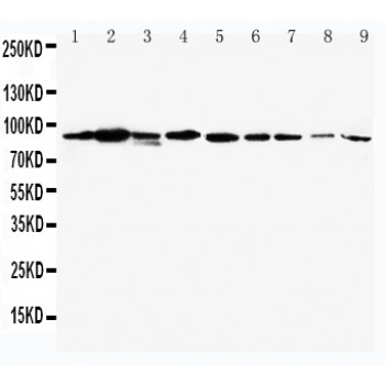
Anti-STAT1 antibody, ASA-B1789, Western blotting
Lane 1: Mouse Heart Tissue Lysate
Lane 2: Mouse Liver Tissue Lysate
Lane 3: Mouse Brain Tissue Lysate
Lane 4: Mouse Kidney Tissue Lysate
Lane 5: Mouse Spleen Tissue Lysate
Lane 6: Mouse Thymus Tissue Lysate
Lane 7: Mouse Lung Tissue Lysate
Lane 8: Mouse Intestine Tissue Lysate
Lane 9: Mouse Ovary Tissue Lysate