More info
Overview
Long Name | Antibody Type | Antibody Isotype | Host | Species Reactivity | Validated Applications | Purification |
| interleukin-1 receptor-associated kinase 1 | Polyclonal | IgG | Rabbit | Human, Mouse, Rat | WB | Immunogen affinity purified. |
Immunogen | ||||||
| A synthetic peptide corresponding to a sequence at the N-terminus of human IRAK(18-37aa FLYEVPPWVMCRFYKVMDAL), identical to the related rat and mouse sequences. | ||||||
Properties
Form | Lyophilized |
Size | 100 µg/vial |
Contents | Antibody is lyophilized with 5 mg BSA, 0.9 mg NaCl, 0.2 mg Na2HPO4, 0.05 mg Thimerosal and 0.05 mg NaN3. *carrier free antibody available upon request. |
Concentration | Reconstitute with 0.2 mL sterile dH2O (500 µg/ml final concentration). |
Storage | At -20 °C for 12 months, as supplied. Store reconstituted antibody at 2-8 °C for one month. For long-term storage, aliquot and store at -20 °C. Avoid repeated freezing and thawing. |
Additional Information Regarding the Antigen
Gene | IRAK1 |
Protein | Interleukin-1 receptor-associated kinase 1 |
Uniprot ID | P51617 |
Function | Serine/threonine-protein kinase that plays a critical role in initiating innate immune response against foreign pathogens. Involved in Toll-like receptor (TLR) and IL-1R signaling pathways. Is rapidly recruited by MYD88 to the receptor- signaling complex upon TLR activation. Association with MYD88 leads to IRAK1 phosphorylation by IRAK4 and subsequent autophosphorylation and kinase activation. Phosphorylates E3 ubiquitin ligases Pellino proteins (PELI1, PELI2 and PELI3) to promote pellino-mediated polyubiquitination of IRAK1. Then, the ubiquitin-binding domain of IKBKG/NEMO binds to polyubiquitinated IRAK1 bringing together the IRAK1-MAP3K7/TAK1-TRAF6 complex and the NEMO-IKKA-IKKB complex. In turn, MAP3K7/TAK1 activates IKKs (CHUK/IKKA and IKBKB/IKKB) leading to NF-kappa-B nuclear translocation and activation. Alternatively, phosphorylates TIRAP to promote its ubiquitination and subsequent degradation. Phosphorylates the interferon regulatory factor 7 (IRF7) to induce its activation and translocation to the nucleus, resulting in transcriptional activation of type I IFN genes, which drive the cell in an antiviral state. When sumoylated, translocates to the nucleus and phosphorylates STAT3. |
Tissue Specificity | Isoform 1 and isoform 2 are ubiquitously expressed in all tissues examined, with isoform 1 being more strongly expressed than isoform 2. |
Sub-cellular localization | Cytoplasm. |
Sequence Similarities | Belongs to the protein kinase superfamily. TKL Ser/Thr protein kinase family. Pelle subfamily. |
Aliases | Il1rak antibody|Interleukin 1 receptor associated kinase 1 antibody|Interleukin-1 receptor-associated kinase 1 antibody|IRAK antibody|IRAK-1 antibody|Irak1 antibody|IRAK1_HUMAN antibody|mPLK antibody|OTTHUMP00000026014 antibody|OTTHUMP00000026015 antibody|OTTHUMP00000026020 antibody|OTTHUMP00000180621 antibody|Pelle antibody|Pelle homolog antibody|Pelle-like protein kinase antibody|Plpk antibody |
Application Details
| Application | Concentration* | Species | Validated Using** |
| Western blot | 0.1-0.5μg/ml | Human, Rat Mouse | AssaySolutio's ECL kit |
AssaySolution recommends Rabbit Chemiluminescent WB Detection Kit (AKIT001B) for Western blot. *Blocking peptide can be purchased at $65. Contact us for more information
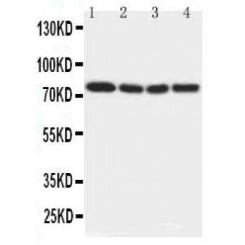
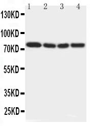
Anti-IRAK antibody, ASA-B1061, Western blotting
Lane 1: Rat Liver Tissue Lysate
Lane 2: Human Placenta Tissue Lysate
Lane 3: MCF-7 Cell Lysate
Lane 4: PANC Cell Lysate
Lane 1: Rat Liver Tissue Lysate
Lane 2: Human Placenta Tissue Lysate
Lane 3: MCF-7 Cell Lysate
Lane 4: PANC Cell Lysate