More info
Overview
Long Name | Antibody Type | Antibody Isotype | Host | Species Reactivity | Validated Applications | Purification |
| melanoma cell adhesion molecule | Polyclonal | IgG | Rabbit | Human, Mouse, Rat | IHC-P, WB | Immunogen affinity purified. |
Immunogen | ||||||
| A synthetic peptide corresponding to a sequence at the C-terminus of human CD146(584-600aa KKGKLPCRRSGKQEITL), different from the related rat and mouse sequences by one amino acid. | ||||||
Properties
Form | Lyophilized |
Size | 100 µg/vial |
Contents | Antibody is lyophilized with 5 mg BSA, 0.9 mg NaCl, 0.2 mg Na2HPO4, 0.05 mg Thimerosal and 0.05 mg NaN3. *carrier free antibody available upon request. |
Concentration | Reconstitute with 0.2 mL sterile dH2O (500 µg/ml final concentration). |
Storage | At -20 °C for 12 months, as supplied. Store reconstituted antibody at 2-8 °C for one month. For long-term storage, aliquot and store at -20 °C. Avoid repeated freezing and thawing. |
Additional Information Regarding the Antigen
Gene | MCAM |
Protein | Cell surface glycoprotein MUC18 |
Uniprot ID | P43121 |
Function | Plays a role in cell adhesion, and in cohesion of the endothelial monolayer at intercellular junctions in vascular tissue. Its expression may allow melanoma cells to interact with cellular elements of the vascular system, thereby enhancing hematogeneous tumor spread. Could be an adhesion molecule active in neural crest cells during embryonic development. Acts as surface receptor that triggers tyrosine phosphorylation of FYN and PTK2/FAK1, and a transient increase in the intracellular calcium concentration. |
Tissue Specificity | Detected in endothelial cells in vascular tissue throughout the body. May appear at the surface of neural crest cells during their embryonic migration. Appears to be limited to vascular smooth muscle in normal adult tissues. Associated with tumor progression and the development of metastasis in human malignant melanoma. Expressed most strongly on metastatic lesions and advanced primary tumors and is only rarely detected in benign melanocytic nevi and thin primary melanomas with a low probability of metastasis. |
Sub-cellular localization | Membrane; Single-pass type I membrane protein. |
Sequence Similarities | Contains 3 Ig-like C2-type (immunoglobulin-like) domains. |
Aliases | A32 antigen antibody|CD 146 antibody|CD146 antibody|CD146 antigen antibody|Cell surface glycoprotein MUC18 antibody|Cell surface glycoprotein P1H12 antibody|Gicerin antibody|MCAM antibody|Melanoma adhesion molecule antibody|Melanoma associated antigen A32 antibody|Melanoma associated antigen MUC18 antibody|Melanoma associated glycoprotein MUC18 antibody|Melanoma cell adhesion molecule antibody|Melanoma-associated antigen A32 antibody|Melanoma-associated antigen MUC18 antibody|MelCAM antibody|MUC 18 antibody|MUC18 antibody|MUC18_HUMAN antibody|S endo 1 antibody|S endo 1 endothelial associated antigen antibody|S-endo 1 endothelial-associated antigen antibody|Sendo 1 endothelial associated antigen antibody|Sendo1 antibody |
Application Details
| Application | Concentration* | Species | Validated Using** |
| Western blot | 0.1-0.5μg/ml | Human Mouse, Rat | AssaySolutio's ECL kit |
| Immunohistochemistry(Paraffin-embedded Section) | 0.5-1μg/ml | Human Mouse, Rat | AssaySolutio's IHC/ICC Detection kit |
AssaySolution recommends Rabbit Chemiluminescent WB Detection Kit (AKIT001B) for Western blot, and Rabbit Peroxidase IHC/ICC Detection Kit (AKIT002B) for IHC(P). *Blocking peptide can be purchased at $65. Contact us for more information
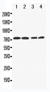
Anti-CD146 antibody, ASA-B0351, Western blotting
Lane 1: A549 Cell Lysate
Lane 2: A549 Cell Lysate
Lane 3: HELA Cell Lysate
Lane 4: SW620 Cell Lysate
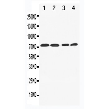
Lane 1: A549 Cell Lysate
Lane 2: A549 Cell Lysate
Lane 3: HELA Cell Lysate
Lane 4: SW620 Cell Lysate
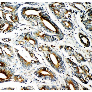
Anti-CD146 antibody, ASA-B0351, IHC(P)
IHC(P): Human Intestinal Cancer Tissue
IHC(P): Human Intestinal Cancer Tissue