More info
Overview
Long Name | Antibody Type | Antibody Isotype | Host | Species Reactivity | Validated Applications | Purification |
| Pim-1 proto-oncogene, serine/threonine kinase | Polyclonal | IgG | Rabbit | Human | WB | Immunogen affinity purified. |
Immunogen | ||||||
| A synthetic peptide corresponding to a sequence at the C-terminus of human PIM1(373-404aa EEIQNHPWMQDVLLPQETAEIHLHSLSPGPSK), different from the related mouse sequence by seven amino acids and from the related rat sequence by two amino acids. | ||||||
Properties
Form | Lyophilized |
Size | 100 µg/vial |
Contents | Antibody is lyophilized with 5 mg BSA, 0.9 mg NaCl, 0.2 mg Na2HPO4, 0.05 mg NaN3. *carrier free antibody available upon request. |
Concentration | Reconstitute with 0.2 mL sterile dH2O (500 µg/ml final concentration). |
Storage | At -20 °C for 12 months, as supplied. Store reconstituted antibody at 2-8 °C for one month. For long-term storage, aliquot and store at -20 °C. Avoid repeated freezing and thawing. |
Additional Information Regarding the Antigen
Gene | PIM1 |
Protein | Serine/threonine-protein kinase pim-1 |
Uniprot ID | P11309 |
Function | Proto-oncogene with serine/threonine kinase activity involved in cell survival and cell proliferation and thus providing a selective advantage in tumorigenesis. Exerts its oncogenic activity through: the regulation of MYC transcriptional activity, the regulation of cell cycle progression and by phosphorylation and inhibition of proapoptotic proteins (BAD, MAP3K5, FOXO3). Phosphorylation of MYC leads to an increase of MYC protein stability and thereby an increase of transcriptional activity. The stabilization of MYC exerted by PIM1 might explain partly the strong synergism between these two oncogenes in tumorigenesis. Mediates survival signaling through phosphorylation of BAD, which induces release of the anti-apoptotic protein Bcl- X(L)/BCL2L1. Phosphorylation of MAP3K5, an other proapoptotic protein, by PIM1, significantly decreases MAP3K5 kinase activity and inhibits MAP3K5-mediated phosphorylation of JNK and JNK/p38MAPK subsequently reducing caspase-3 activation and cell apoptosis. Stimulates cell cycle progression at the G1-S and G2-M transitions by phosphorylation of CDC25A and CDC25C. Phosphorylation of CDKN1A, a regulator of cell cycle progression at G1, results in the relocation of CDKN1A to the cytoplasm and enhanced CDKN1A protein stability. Promote cell cycle progression and tumorigenesis by down-regulating expression of a regulator of cell cycle progression, CDKN1B, at both transcriptional and post- translational levels. Phosphorylation of CDKN1B,induces 14-3-3- proteins binding, nuclear export and proteasome-dependent degradation. May affect the structure or silencing of chromatin by phosphorylating HP1 gamma/CBX3. Acts also as a regulator of homing and migration of bone marrow cells involving functional interaction with the CXCL12-CXCR4 signaling axis. |
Tissue Specificity | Expressed primarily in cells of the hematopoietic and germline lineages. Isoform 1 and isoform 2 are both expressed in prostate cancer cell lines. |
Sub-cellular localization | Isoform 2: Cytoplasm. Nucleus. |
Sequence Similarities | Belongs to the protein kinase superfamily. CAMK Ser/Thr protein kinase family. PIM subfamily. |
Aliases | Oncogene PIM 1 antibody|Oncogene PIM1 antibody|PIM 1 antibody|pim 1 kinase 44 kDa isoform antibody|Pim 1 kinase antibody|pim 1 oncogene (proviral integration site 1) antibody|Pim 1 oncogene antibody|PIM antibody|PIM1 antibody|pim1 kinase 44 kDa isoform antibody|PIM1_HUMAN antibody|Pim2 antibody|PIM3 antibody|Proto oncogene serine/threonine protein kinase Pim 1 antibody|Proto-oncogene serine/threonine-protein kinase Pim-1 antibody|Proviral integration site 1 antibody|Proviral integration site 2 antibody |
Application Details
| Application | Concentration* | Species | Validated Using** |
| Western blot | 0.1-0.5μg/ml | Human | AssaySolutio's ECL kit |
AssaySolution recommends Rabbit Chemiluminescent WB Detection Kit (AKIT001B) for Western blot. *Blocking peptide can be purchased at $65. Contact us for more information

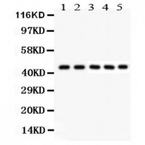
Anti- PIM1 antibody, ASA-B1523, Western blotting
All lanes: Anti PIM1 (ASA-B1523) at 0.5ug/ml
Lane 1: U20S Whole Cell Lysate at 40ug
Lane 2: A549 Whole Cell Lysate at 40ug
Lane 3: COLO320 Whole Cell Lysate at 40ug
Lane 4: SW620 Whole Cell Lysate at 40ug
Lane 5: JURKAT Whole Cell Lysate at 40ug
Predicted bind size: 45KD
Observed bind size: 45KD
All lanes: Anti PIM1 (ASA-B1523) at 0.5ug/ml
Lane 1: U20S Whole Cell Lysate at 40ug
Lane 2: A549 Whole Cell Lysate at 40ug
Lane 3: COLO320 Whole Cell Lysate at 40ug
Lane 4: SW620 Whole Cell Lysate at 40ug
Lane 5: JURKAT Whole Cell Lysate at 40ug
Predicted bind size: 45KD
Observed bind size: 45KD


