More info
Overview
Long Name | Antibody Type | Antibody Isotype | Host | Species Reactivity | Validated Applications | Purification |
| autophagy related 5 | Polyclonal | IgG | Rabbit | Human, Mouse, Rat | WB | Immunogen affinity purified. |
Immunogen | ||||||
| A synthetic peptide corresponding to a sequence in the middle region of human APG5L(82-97aa DRFDQFWAINRKLMEY), identical to the related mouse sequence, and different from the related rat sequence by one amino acid. | ||||||
Properties
Form | Lyophilized |
Size | 100 µg/vial |
Contents | Antibody is lyophilized with 5 mg BSA, 0.9 mg NaCl, 0.2 mg Na2HPO4, 0.05 mg Thimerosal and 0.05 mg NaN3. *carrier free antibody available upon request. |
Concentration | Reconstitute with 0.2 mL sterile dH2O (500 µg/ml final concentration). |
Storage | At -20 °C for 12 months, as supplied. Store reconstituted antibody at 2-8 °C for one month. For long-term storage, aliquot and store at -20 °C. Avoid repeated freezing and thawing. |
Additional Information Regarding the Antigen
Gene | ATG5 |
Protein | Autophagy protein 5 |
Uniprot ID | Q9H1Y0 |
Function | Involved in autophagic vesicle formation. Conjugation with ATG12, through a ubiquitin-like conjugating system involving ATG7 as an E1-like activating enzyme and ATG10 as an E2-like conjugating enzyme, is essential for its function. The ATG12-ATG5 conjugate acts as an E3-like enzyme which is required for lipidation of ATG8 family proteins and their association to the vesicle membranes. Involved in mitochondrial quality control after oxidative damage, and in subsequent cellular longevity. The ATG12- ATG5 conjugate also negatively regulates the innate antiviral immune response by blocking the type I IFN production pathway through direct association with RARRES3 and MAVS. Also plays a role in translation or delivery of incoming viral RNA to the translation apparatus. Plays a critical role in multiple aspects of lymphocyte development and is essential for both B and T lymphocyte survival and proliferation. Required for optimal processing and presentation of antigens for MHC II. Involved in the maintenance of axon morphology and membrane structures, as well as in normal adipocyte differentiation. Promotes primary ciliogenesis through removal of OFD1 from centriolar satellites and degradation of IFT20 via the autophagic pathway. |
Tissue Specificity | Ubiquitous. The mRNA is present at similar levels in viable and apoptotic cells, whereas the protein is dramatically highly expressed in apoptotic cells. |
Sub-cellular localization | Cytoplasm. Preautophagosomal structure membrane; Peripheral membrane protein. Note: Colocalizes with nonmuscle actin. The conjugate detaches from the membrane immediately before or after autophagosome formation is completed (By similarity). Localizes also to discrete punctae along the ciliary axoneme and to the base of the ciliary axoneme. |
Sequence Similarities | |
Aliases | APG 5 antibody|APG 5L antibody|APG5 antibody|APG5 autophagy 5 like antibody|APG5 like antibody|APG5-like antibody|APG5L antibody|Apoptosis specific protein antibody|Apoptosis-specific protein antibody|ASP antibody|ATG 5 antibody|Atg5 antibody|ATG5 autophagy related 5 homolog antibody|Autophagy protein 5 antibody|hAPG5 antibody|Homolog of S Cerevisiae autophagy 5 antibody|OTTHUMP00000040507 antibody |
Application Details
| Application | Concentration* | Species | Validated Using** |
| Western blot | 0.1-0.5μg/ml | Human, Mouse, Rat | AssaySolutio's ECL kit |
AssaySolution recommends Rabbit Chemiluminescent WB Detection Kit (AKIT001B) for Western blot. *Blocking peptide can be purchased at $65. Contact us for more information

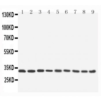
Anti-APG5L/ATG5 antibody, ASA-B0113, All Western blotting
All lanes: Anti- ATG5(ASA-B0113) at 0.5ug/ml
Lane 1: Rat Liver Tissue Lysate at 4oug
Lane 2: Rat Spleen Tissue Lysate at 4oug
Lane 3: Rat Kidney Tissue Lysate at 4oug
Lane 4: HELA Whole Cell Lysate at 40ug
Lane 5: RAJI Whole Cell Lysate at 40ug
Lane 6: NIH Whole Cell Lysate at 40ug
Lane 7: HEPG2 Whole Cell Lysate at 40ug
Lane 8: PC12 Whole Cell Lysate at 40ug
Lane 9: NRK Whole Cell Lysate at 40ug


