

More info
Overview
Long Name | Antibody Type | Antibody Isotype | Host | Species Reactivity | Validated Applications | Purification |
| TGF-beta activated kinase 1/MAP3K7 binding protein 2 | Polyclonal | IgG | Rabbit | Human, Mouse, Rat | IHC-P, WB | Immunogen affinity purified. |
Immunogen | ||||||
| A synthetic peptide corresponding to a sequence at the C-terminus of human TAB2(487-506aa LTNLLNHPDHYVETENIQHL), identical to the related mouse sequence, and different from the related rat sequence by one amino acid. | ||||||
Properties
Form | Lyophilized |
Size | 100 µg/vial |
Contents | Antibody is lyophilized with 5 mg BSA, 0.9 mg NaCl, 0.2 mg Na2HPO4, 0.05 mg Thimerosal and 0.05 mg NaN3. *carrier free antibody available upon request. |
Concentration | Reconstitute with 0.2 mL sterile dH2O (500 µg/ml final concentration). |
Storage | At -20 °C for 12 months, as supplied. Store reconstituted antibody at 2-8 °C for one month. For long-term storage, aliquot and store at -20 °C. Avoid repeated freezing and thawing. |
Additional Information Regarding the Antigen
Gene | TAB2 |
Protein | TGF-beta-activated kinase 1/MAP3K7-binding protein 2 |
Uniprot ID | Q9NYJ8 |
Function | Adapter linking MAP3K7/TAK1 and TRAF6. Promotes MAP3K7 activation in the IL1 signaling pathway. The binding of 'Lys-63'- linked polyubiquitin chains to TAB2 promotes autophosphorylation of MAP3K7 at 'Thr-187'. Involved in heart development. |
Tissue Specificity | Widely expressed. In the embryo, expressed in the ventricular trabeculae, endothelial cells of the conotruncal cushions of the outflow tract and in the endothelial cells lining the developing aortic valves. |
Sub-cellular localization | Membrane. |
Sequence Similarities | Contains 1 CUE domain. |
Aliases | CHTD2 antibody|FLJ21885 antibody|KIAA0733 antibody|MAP3K7IP2 antibody|Mitogen activated protein kinase kinase kinase 7 interacting protein 2 antibody|Mitogen-activated protein kinase kinase kinase 7-interacting protein 2 antibody|OTTHUMP00000040125 antibody|TAB 2 antibody|TAB-2 antibody|Tab2 antibody|TAK1 binding protein 2 antibody|TAK1-binding protein 2 antibody|TGF beta activated kinase 1/MAP3K7 binding protein 2 antibody|TGF-beta-activated kinase 1 and MAP3K7-binding protein 2 antibody|TGF-beta-activated kinase 1-binding protein 2 antibody |
Application Details
| Application | Concentration* | Species | Validated Using** |
| Western blot | 0.1-0.5μg/ml | Human, Mouse, Rat | AssaySolutio's ECL kit |
| Immunohistochemistry(Paraffin-embedded Section) | 0.5-1μg/ml | Human Mouse, Rat | AssaySolutio's IHC/ICC Detection kit |
AssaySolution recommends Rabbit Chemiluminescent WB Detection Kit (AKIT001B) for Western blot, and Rabbit Peroxidase IHC/ICC Detection Kit (AKIT002B) for IHC(P). *Blocking peptide can be purchased at $65. Contact us for more information
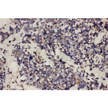
Anti-TAB2 antibody, ASA-B1829, IHC(P)
IHC(P): Human Lung Cancer Tissue
IHC(P): Human Lung Cancer Tissue
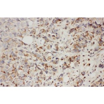
Anti-TAB2 antibody, ASA-B1829, IHC(P)
IHC(P): Human Mammary Cancer Tissue
IHC(P): Human Mammary Cancer Tissue
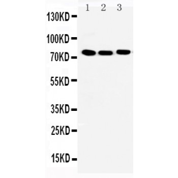
Anti-TAB2 antibody, ASA-B1829, All Western blotting
All lanes: Anti-TAB2(ASA-B1829) at 0.5ug/ml
Lane 1: Mouse Brain Tissue Lysate at 40ug
Lane 2: Mouse Testis Tissue Lysate at 40ug
Lane 3: Mouse Spleen Tissue Lysate at 40ug
Predicted bind size: 76KD
Observed bind size: 76KD
All lanes: Anti-TAB2(ASA-B1829) at 0.5ug/ml
Lane 1: Mouse Brain Tissue Lysate at 40ug
Lane 2: Mouse Testis Tissue Lysate at 40ug
Lane 3: Mouse Spleen Tissue Lysate at 40ug
Predicted bind size: 76KD
Observed bind size: 76KD