More info
Overview
Long Name | Antibody Type | Antibody Isotype | Host | Species Reactivity | Validated Applications | Purification |
| protein kinase C, iota | Polyclonal | IgG | Rabbit | Human, Mouse, Rat | IHC-P, WB | Immunogen affinity purified. |
Immunogen | ||||||
| E.coli-derived human PKC iota recombinant protein (Position: D21-Q214). Human PKC iota shares 96% and 97% amino acid (aa) sequence identity with mouse and rat PKC iota, respectively. | ||||||
Properties
Form | Lyophilized |
Size | 100 µg/vial |
Contents | Antibody is lyophilized with 5 mg BSA, 0.9 mg NaCl, 0.2 mg Na2HPO4, 0.05 mg NaN3. *carrier free antibody available upon request. |
Concentration | Reconstitute with 0.2 mL sterile dH2O (500 µg/ml final concentration). |
Storage | At -20 °C for 12 months, as supplied. Store reconstituted antibody at 2-8 °C for one month. For long-term storage, aliquot and store at -20 °C. Avoid repeated freezing and thawing. |
Additional Information Regarding the Antigen
Gene | PRKCI |
Protein | Protein kinase C iota type |
Uniprot ID | P41743 |
Function | Calcium- and diacylglycerol-independent serine/ threonine-protein kinase that plays a general protective role against apoptotic stimuli, is involved in NF-kappa-B activation, cell survival, differentiation and polarity, and contributes to the regulation of microtubule dynamics in the early secretory pathway. Is necessary for BCR-ABL oncogene-mediated resistance to apoptotic drug in leukemia cells, protecting leukemia cells against drug-induced apoptosis. In cultured neurons, prevents amyloid beta protein-induced apoptosis by interrupting cell death process at a very early step. In glioblastoma cells, may function downstream of phosphatidylinositol 3-kinase (PI(3)K) and PDPK1 in the promotion of cell survival by phosphorylating and inhibiting the pro-apoptotic factor BAD. Can form a protein complex in non- small cell lung cancer (NSCLC) cells with PARD6A and ECT2 and regulate ECT2 oncogenic activity by phosphorylation, which in turn promotes transformed growth and invasion. In response to nerve growth factor (NGF), acts downstream of SRC to phosphorylate and activate IRAK1, allowing the subsequent activation of NF-kappa-B and neuronal cell survival. Functions in the organization of the apical domain in epithelial cells by phosphorylating EZR. This step is crucial for activation and normal distribution of EZR at the early stages of intestinal epithelial cell differentiation. Forms a protein complex with LLGL1 and PARD6B independently of PARD3 to regulate epithelial cell polarity. Plays a role in microtubule dynamics in the early secretory pathway through interaction with RAB2A and GAPDH and recruitment to vesicular tubular clusters (VTCs). In human coronary artery endothelial cells (HCAEC), is activated by saturated fatty acids and mediates lipid-induced apoptosis. |
Tissue Specificity | Predominantly expressed in lung and brain, but also expressed at lower levels in many tissues including pancreatic islets. Highly expressed in non-small cell lung cancers. |
Sub-cellular localization | Cytoplasm. Membrane. Endosome. Nucleus. Note: Transported into the endosome through interaction with SQSTM1/p62. After phosphorylation by SRC, transported into the nucleus through interaction with KPNB1. Colocalizes with CDK7 in the cytoplasm and nucleus. Transported to vesicular tubular clusters (VTCs) through interaction with RAB2A. |
Sequence Similarities | Belongs to the protein kinase superfamily. AGC Ser/Thr protein kinase family. PKC subfamily. |
Aliases | aPKC lambda/iota antibody|aPKC-lambda/iota antibody|Atypical protein kinase C lambda/iota antibody|Atypical protein kinase C-lambda/iota antibody|DXS1179E antibody|KPCI_HUMAN antibody|MGC26534 antibody|nPKC iota antibody|nPKC-iota antibody|nPKCiota antibody|PKC lambda antibody|PKCI antibody|PKCiota antibody|PKClambda antibody|PRKC I antibody|PRKC iota antibody|PRKC lambda/iota antibody|PRKC-lambda/iota antibody|PRKCI antibody|Protein kinase C iota antibody|Protein kinase C iota type antibody |
Application Details
| Application | Concentration* | Species | Validated Using** |
| Western blot | 0.1-0.5μg/ml | Human, Mouse, Rat | AssaySolutio's ECL kit |
| Immunohistochemistry(Paraffin-embedded Section) | 0.5-1μg/ml | Human, Mouse, Rat | AssaySolutio's IHC/ICC Detection kit |
AssaySolution recommends Rabbit Chemiluminescent WB Detection Kit (AKIT001B) for Western blot, and Rabbit Peroxidase IHC/ICC Detection Kit (AKIT002B) for IHC(P). *Blocking peptide can be purchased at $65. Contact us for more information

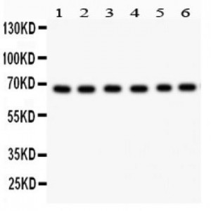
Anti- PKC iota antibody, ASA-B1534, Western blotting
All lanes: Anti PKC iota (ASA-B1534) at 0.5ug/ml
Lane 1: SHG Whole Cell Lysate at 40ug
Lane 2: A549 Whole Cell Lysate at 40ug
Lane 3: U87 Whole Cell Lysate at 40ug
Lane 4: 293T Whole Cell Lysate at 40ug
Lane 5: HELA Whole Cell Lysate at 40ug
Lane 6: JURKAT Whole Cell Lysate at 40ug
Predicted bind size: 68KD
Observed bind size: 68KD

Anti- PKC iota antibody, ASA-B1534,IHC(P)
IHC(P): Mouse Brain Tissue
IHC(P): Mouse Brain Tissue

Anti- PKC iota antibody, ASA-B1534,IHC(P)
IHC(P): Rat Brain Tissue
IHC(P): Rat Brain Tissue


