More info
Overview
Long Name | Antibody Type | Antibody Isotype | Host | Species Reactivity | Validated Applications | Purification |
| tumor protein p53 | Polyclonal | IgG | Rabbit | Human | IHC-P, WB | Immunogen affinity purified. |
Immunogen | ||||||
| E.coli-derived human P53 recombinant protein (Position: A74-D393). Human P53 shares 83% and 85% amino acid (aa) sequences identity with mouse and rat P53, respectively. | ||||||
Properties
Form | Lyophilized |
Size | 100 µg/vial |
Contents | Antibody is lyophilized with 5 mg BSA, 0.9 mg NaCl, 0.2 mg Na2HPO4, 0.05 mg NaN3. *carrier free antibody available upon request. |
Concentration | Reconstitute with 0.2 mL sterile dH2O (500 µg/ml final concentration). |
Storage | At -20 °C for 12 months, as supplied. Store reconstituted antibody at 2-8 °C for one month. For long-term storage, aliquot and store at -20 °C. Avoid repeated freezing and thawing. |
Additional Information Regarding the Antigen
Gene | TP53 |
Protein | Cellular tumor antigen p53 |
Uniprot ID | P04637 |
Function | Acts as a tumor suppressor in many tumor types; induces growth arrest or apoptosis depending on the physiological circumstances and cell type. Involved in cell cycle regulation as a trans-activator that acts to negatively regulate cell division by controlling a set of genes required for this process. One of the activated genes is an inhibitor of cyclin-dependent kinases. Apoptosis induction seems to be mediated either by stimulation of BAX and FAS antigen expression, or by repression of Bcl-2 expression. In cooperation with mitochondrial PPIF is involved in activating oxidative stress-induced necrosis; the function is largely independent of transcription. Induces the transcription of long intergenic non-coding RNA p21 (lincRNA-p21) and lincRNA- Mkln1. LincRNA-p21 participates in TP53-dependent transcriptional repression leading to apoptosis and seem to have to effect on cell-cycle regulation. Implicated in Notch signaling cross-over. Prevents CDK7 kinase activity when associated to CAK complex in response to DNA damage, thus stopping cell cycle progression. Isoform 2 enhances the transactivation activity of isoform 1 from some but not all TP53-inducible promoters. Isoform 4 suppresses transactivation activity and impairs growth suppression mediated by isoform 1. Isoform 7 inhibits isoform 1-mediated apoptosis. Regulates the circadian clock by repressing CLOCK-ARNTL/BMAL1- mediated transcriptional activation of PER2 (PubMed:24051492). |
Tissue Specificity | Ubiquitous. Isoforms are expressed in a wide range of normal tissues but in a tissue-dependent manner. Isoform 2 is expressed in most normal tissues but is not detected in brain, lung, prostate, muscle, fetal brain, spinal cord and fetal liver. Isoform 3 is expressed in most normal tissues but is not detected in lung, spleen, testis, fetal brain, spinal cord and fetal liver. Isoform 7 is expressed in most normal tissues but is not detected in prostate, uterus, skeletal muscle and breast. Isoform 8 is detected only in colon, bone marrow, testis, fetal brain and intestine. Isoform 9 is expressed in most normal tissues but is not detected in brain, heart, lung, fetal liver, salivary gland, breast or intestine. |
Sub-cellular localization | Cytoplasm. Nucleus. Nucleus, PML body. Endoplasmic reticulum. Mitochondrion matrix. Note: Interaction with BANP promotes nuclear localization. Recruited into PML bodies together with CHEK2. Translocates to mitochondria upon oxidative stress. |
Sequence Similarities | Belongs to the p53 family. |
Aliases | Antigen NY-CO-13 antibody|BCC7 antibody|Cellular tumor antigen p53 antibody|Cys 51 Stop antibody|FLJ92943 antibody|HGNC11998 antibody|LFS1 antibody|Mutant tumor protein 53 antibody|p53 antibody|p53 Cellular Tumor Antigen antibody|p53 tumor suppressor antibody|P53_HUMAN antibody|Phosphoprotein p53 antibody|Tp53 antibody|Transformation related protein 53 antibody|TRP53 antibody|Tumor protein p53 antibody|Tumor suppressor p53 antibody|Tumour Protein p53 antibody |
Application Details
| Application | Concentration* | Species | Validated Using** |
| Western blot | 0.1-0.5μg/ml | Human | AssaySolutio's ECL kit |
| Immunohistochemistry(Paraffin-embedded Section) | 0.5-1μg/ml | Human | AssaySolutio's IHC/ICC Detection kit |
AssaySolution recommends Rabbit Chemiluminescent WB Detection Kit (AKIT001B) for Western blot, and Rabbit Peroxidase IHC/ICC Detection Kit (AKIT002B) for IHC(P). *Blocking peptide can be purchased at $65. Contact us for more information

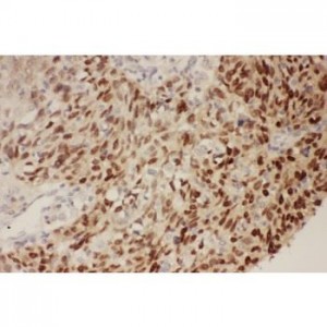
Anti-P53 antibody, ASA-B1461--1.jpg
IHC(P): Human Lung Cancer Tissue
IHC(P): Human Lung Cancer Tissue

Anti-P53 antibody, ASA-B1461--2.jpg
All lanes: Anti-P53(ASA-B1461) at 0.5ug/ml
Lane 1: HEPG2 Whole Cell Lysate at 40ug
Lane 2: COLO320 Whole Cell Lysate at 40ug
Predicted bind size: 53KD
Observed bind size: 53KD
All lanes: Anti-P53(ASA-B1461) at 0.5ug/ml
Lane 1: HEPG2 Whole Cell Lysate at 40ug
Lane 2: COLO320 Whole Cell Lysate at 40ug
Predicted bind size: 53KD
Observed bind size: 53KD


