More info
Overview
Long Name | Antibody Type | Antibody Isotype | Host | Species Reactivity | Validated Applications | Purification |
| optineurin | Polyclonal | IgG | Rabbit | Human | IHC-P, WB | Immunogen affinity purified. |
Immunogen | ||||||
| E.coli-derived human Optineurin recombinant protein (Position: R241-I577). Human Optineurin shares 82% amino acid (aa) sequence identity with both mouse and rat Optineurin. | ||||||
Properties
Form | Lyophilized |
Size | 100 µg/vial |
Contents | Antibody is lyophilized with 5 mg BSA, 0.9 mg NaCl, 0.2 mg Na2HPO4, 0.05 mg NaN3. *carrier free antibody available upon request. |
Concentration | Reconstitute with 0.2 mL sterile dH2O (500 µg/ml final concentration). |
Storage | At -20 °C for 12 months, as supplied. Store reconstituted antibody at 2-8 °C for one month. For long-term storage, aliquot and store at -20 °C. Avoid repeated freezing and thawing. |
Additional Information Regarding the Antigen
Gene | OPTN |
Protein | Optineurin |
Uniprot ID | Q96CV9 |
Function | Plays an important role in the maintenance of the Golgi complex, in membrane trafficking, in exocytosis, through its interaction with myosin VI and Rab8. Links myosin VI to the Golgi complex and plays an important role in Golgi ribbon formation. Negatively regulates the induction of IFNB in response to RNA virus infection. Plays a neuroprotective role in the eye and optic nerve. Probably part of the TNF-alpha signaling pathway that can shift the equilibrium toward induction of cell death. May act by regulating membrane trafficking and cellular morphogenesis via a complex that contains Rab8 and hungtingtin (HD). Mediates the interaction of Rab8 with the probable GTPase-activating protein TBC1D17 during Rab8-mediated endocytic trafficking, such as of transferrin receptor (TFRC/TfR); regulates Rab8 recruitnment to tubules emanating from the endocytic recycling compartment. Autophagy receptor that interacts directly with both the cargo to become degraded and an autophagy modifier of the MAP1 LC3 family; targets ubiquitin-coated bacteria (xenophagy), such as cytoplasmic Salmonella enterica, and appears to function in the same pathway as SQSTM1 and CALCOCO2/NDP52. May constitute a cellular target for adenovirus E3 14.7, an inhibitor of TNF-alpha functions, thereby affecting cell death. |
Tissue Specificity | Present in aqueous humor of the eye (at protein level). Highly expressed in trabecular meshwork. Expressed nonpigmented ciliary epithelium, retina, brain, adrenal cortex, fetus, lymphocyte, fibroblast, skeletal muscle, heart, liver, brain and placenta. |
Sub-cellular localization | Cytoplasm, perinuclear region. Golgi apparatus. Golgi apparatus, trans-Golgi network. Cytoplasmic vesicle, autophagosome. Cytoplasmic vesicle. Recycling endosome. Note: Found in the perinuclear region and associates with the Golgi apparatus. Colocalizes with MYO6 and RAB8 at the Golgi complex and in vesicular structures close to the plasma membrane. Localizes to LC3-positive cytoplasmic vesicles upon induction of autophagy. |
Sequence Similarities | |
Aliases | 14.7K interacting protein antibody|Ag9 C5 antibody|ALS12 antibody|E3 14.7K interacting protein antibody|E3-14.7K-interacting protein antibody|FIP 2 antibody|FIP-2 antibody|FIP2 antibody|Glaucoma 1 open angle E (adult onset) antibody|Glaucoma 1 open angle E antibody|GLC1E antibody|HIP 7 antibody|HIP-7 antibody|HIP7 antibody|Huntingtin interacting protein 7 antibody|Huntingtin interacting protein HYPL antibody|Huntingtin interacting protein L antibody|Huntingtin yeast partner L antibody|Huntingtin-interacting protein 7 antibody|Huntingtin-interacting protein L antibody|HYPL antibody|Injury inducible protein I 55 antibody|NEMO related protein antibody|NEMO-related protein antibody|NRP antibody|Optic neuropathy inducing protein antibody|Optic neuropathy-inducing protein antibody|Optineurin antibody|OPTN antibody|OPTN_HUMAN antibody|TFIIIA IntP antibody|TFIIIA-IntP antibody|Transcription factor IIIA interacting protein antibody|Transcription factor IIIA-interacting protein antibody|Tumor necrosis factor alpha inducible cellular protein containing leucine zipper domains antibody |
Application Details
| Application | Concentration* | Species | Validated Using** |
| Western blot | 0.1-0.5μg/ml | Human | AssaySolutio's ECL kit |
| Immunohistochemistry(Paraffin-embedded Section) | 0.5-1μg/ml | Human | AssaySolutio's IHC/ICC Detection kit |
AssaySolution recommends Rabbit Chemiluminescent WB Detection Kit (AKIT001B) for Western blot, and Rabbit Peroxidase IHC/ICC Detection Kit (AKIT002B) for IHC(P). *Blocking peptide can be purchased at $65. Contact us for more information

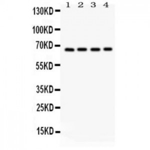
Anti- Optineurin antibody, ASA-B1433, Western blotting
All lanes: Anti Optineurin (ASA-B1433) at 0.5ug/ml
Lane 1: HELA Whole Cell Lysate at 40ug
Lane 2: U87 Whole Cell Lysate at 40ug
Lane 3: SMMC Whole Cell Lysate at 40ug
Lane 4: HT1080 Whole Cell Lysate at 40ug
Predicted bind size: 66KD
Observed bind size: 66KD
All lanes: Anti Optineurin (ASA-B1433) at 0.5ug/ml
Lane 1: HELA Whole Cell Lysate at 40ug
Lane 2: U87 Whole Cell Lysate at 40ug
Lane 3: SMMC Whole Cell Lysate at 40ug
Lane 4: HT1080 Whole Cell Lysate at 40ug
Predicted bind size: 66KD
Observed bind size: 66KD

Anti- Optineurin antibody, ASA-B1433, IHC(P)
IHC(P): Human Placenta Tissue
IHC(P): Human Placenta Tissue


