More info
Overview
Long Name | Antibody Type | Antibody Isotype | Host | Species Reactivity | Validated Applications | Purification |
| nuclear factor of activated T-cells, cytoplasmic, calcineurin-dependent 2 | Polyclonal | IgG | Rabbit | Human, Mouse, Rat | IHC-P, WB | Immunogen affinity purified. |
Immunogen | ||||||
| A synthetic peptide corresponding to a sequence at the C-terminus of human NFAT1(895-913aa KQEQNLDQTYLDDVNEIIR), identical to the related rat and mouse sequences. | ||||||
Properties
Form | Lyophilized |
Size | 100 µg/vial |
Contents | Antibody is lyophilized with 5 mg BSA, 0.9 mg NaCl, 0.2 mg Na2HPO4, 0.05 mg Thimerosal and 0.05 mg NaN3. *carrier free antibody available upon request. |
Concentration | Reconstitute with 0.2 mL sterile dH2O (500 µg/ml final concentration). |
Storage | At -20 °C for 12 months, as supplied. Store reconstituted antibody at 2-8 °C for one month. For long-term storage, aliquot and store at -20 °C. Avoid repeated freezing and thawing. |
Additional Information Regarding the Antigen
Gene | NFATC2 |
Protein | Nuclear factor of activated T-cells, cytoplasmic 2(NF-ATc2/NFATc2) |
Uniprot ID | Q13469 |
Function | Plays a role in the inducible expression of cytokine genes in T-cells, especially in the induction of the IL-2, IL-3, IL-4, TNF-alpha or GM-CSF. Promotes invasive migration through the activation of GPC6 expression and WNT5A signaling pathway. |
Tissue Specificity | Expressed in thymus, spleen, heart, testis, brain, placenta, muscle and pancreas. Isoform 1 is highly expressed in the small intestine, heart, testis, prostate, thymus, placenta and thyroid. Isoform 3 is highly expressed in stomach, uterus, placenta, trachea and thyroid. |
Sub-cellular localization | Cytoplasm. Nucleus. Note: Cytoplasmic for the phosphorylated form and nuclear after activation that is controlled by calcineurin-mediated dephosphorylation. Rapid nuclear exit of NFATC is thought to be one mechanism by which cells distinguish between sustained and transient calcium signals. The subcellular localization of NFATC plays a key role in the regulation of gene transcription. |
Sequence Similarities | Contains 1 RHD (Rel-like) domain. |
Aliases | AI607462 antibody|cytoplasmic 2 antibody|KIAA0611 antibody|NF ATp antibody|NF-ATc2 antibody|NF-ATp antibody|NFAC2_HUMAN antibody|NFAT 1 antibody|NFAT pre existing subunit antibody|NFAT pre-existing subunit antibody|NFAT transcription complex, preexisting component antibody|NFAT1 antibody|NFAT1-D antibody|NFATc2 antibody|NFATp antibody|Nuclear factor of activated T cells cytoplasmic 2 antibody|Nuclear factor of activated T cells cytoplasmic calcineurin dependent 2 antibody|Nuclear factor of activated T cells pre-existing component antibody|Nuclear factor of activated T-cells antibody|T cell transcription factor NFAT 1 antibody|T-cell transcription factor NFAT1 antibody |
Application Details
| Application | Concentration* | Species | Validated Using** |
| Western blot | 0.1-0.5μg/ml | Human, Rat Mouse | AssaySolutio's ECL kit |
| Immunohistochemistry(Paraffin-embedded Section) | 0.5-1μg/ml | Human, Rat Mouse | AssaySolutio's IHC/ICC Detection kit |
AssaySolution recommends Rabbit Chemiluminescent WB Detection Kit (AKIT001B) for Western blot, and Rabbit Peroxidase IHC/ICC Detection Kit (AKIT002B) for IHC(P). *Blocking peptide can be purchased at $65. Contact us for more information

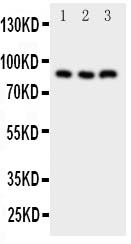
Anti-NFAT1 antibody, ASA-B1370, Western blotting
All lanes: Anti NFAT1 (ASA-B1370) at 0.5ug/ml
Lane 1: PANC Whole Cell Lysate at 40ug
Lane 2: HELA Whole Cell Lysate at 40ug
Lane 3: U87 Whole Cell Lysate at 40ug
Predicted bind size: 87KD
Observed bind size: 87KD
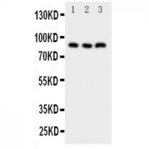
All lanes: Anti NFAT1 (ASA-B1370) at 0.5ug/ml
Lane 1: PANC Whole Cell Lysate at 40ug
Lane 2: HELA Whole Cell Lysate at 40ug
Lane 3: U87 Whole Cell Lysate at 40ug
Predicted bind size: 87KD
Observed bind size: 87KD

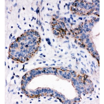
Anti-NFAT1 antibody, ASA-B1370, IHC(P)
IHC(P): Human Mammary Cancer Tissue
IHC(P): Human Mammary Cancer Tissue


