More info
Overview
Long Name | Antibody Type | Antibody Isotype | Host | Species Reactivity | Validated Applications | Purification |
| lamin A/C | Polyclonal | IgG | Rabbit | Human, Mouse, Rat | IHC-P, ICC, WB | Immunogen affinity purified. |
Immunogen | ||||||
| E.coli-derived human Lamin A/C recombinant protein (Position: Y481-Y646). Human Lamin A/C shares 90% and 92% amino acid (aa) sequence identity with mouse and rat Lamin A/C, respectively. | ||||||
Properties
Form | Lyophilized |
Size | 100 µg/vial |
Contents | Antibody is lyophilized with 5 mg BSA, 0.9 mg NaCl, 0.2 mg Na2HPO4, 0.05 mg NaN3. *carrier free antibody available upon request. |
Concentration | Reconstitute with 0.2 mL sterile dH2O (500 µg/ml final concentration). |
Storage | At -20 °C for 12 months, as supplied. Store reconstituted antibody at 2-8 °C for one month. For long-term storage, aliquot and store at -20 °C. Avoid repeated freezing and thawing. |
Additional Information Regarding the Antigen
Gene | LMNA |
Protein | Prelamin-A/C |
Uniprot ID | P02545 |
Function | Lamins are components of the nuclear lamina, a fibrous layer on the nucleoplasmic side of the inner nuclear membrane, which is thought to provide a framework for the nuclear envelope and may also interact with chromatin. Lamin A and C are present in equal amounts in the lamina of mammals. Plays an important role in nuclear assembly, chromatin organization, nuclear membrane and telomere dynamics. Required for normal development of peripheral nervous system and skeletal muscle and for muscle satellite cell proliferation. Required for osteoblastogenesis and bone formation. Also prevents fat infiltration of muscle and bone marrow, helping to maintain the volume and strength of skeletal muscle and bone. |
Tissue Specificity | In the arteries, prelamin-A/C accumulation is not observed in young healthy vessels but is prevalent in medial vascular smooth muscle cells (VSMCs) from aged individuals and in atherosclerotic lesions, where it often colocalizes with senescent and degenerate VSMCs. Prelamin-A/C expression increases with age and disease. In normal aging, the accumulation of prelamin-A/C is caused in part by the down-regulation of ZMPSTE24/FACE1 in response to oxidative stress. |
Sub-cellular localization | Nucleus. Nucleus envelope. Nucleus lamina. Nucleus, nucleoplasm. Note: Farnesylation of prelamin-A/C facilitates nuclear envelope targeting and subsequent cleaveage by ZMPSTE24/FACE1 to remove the farnesyl group produces mature lamin- A/C, which can then be inserted into the nuclear lamina. EMD is required for proper localization of non-farnesylated prelamin-A/C. |
Sequence Similarities | Belongs to the intermediate filament family. |
Aliases | 70 kDa lamin antibody|CDCD1 antibody|CDDC antibody|CMD1A antibody|CMT2B1 antibody|EMD2 antibody|FPL antibody|FPLD antibody|HGPS antibody|IDC antibody|LAMIN A antibody|lamin A/C antibody|LAMIN C antibody|Lamin-A/C antibody|LDP1 antibody|LFP antibody|LGMD1B antibody|LMN 1 antibody|LMN A antibody|LMN C antibody|LMN1 antibody|LMNA antibody|LMNA_HUMAN antibody|LMNC antibody|NY REN 32 antigen antibody|PRO1 antibody|Renal carcinoma antigen NY-REN-32 antibody |
Application Details
| Application | Concentration* | Species | Validated Using** |
| Western blot | 0.1-0.5μg/ml | Human | AssaySolutio's ECL kit |
| Immunohistochemistry(Paraffin-embedded Section) | 0.5-1μg/ml | Human, Mouse, Rat | AssaySolutio's IHC/ICC Detection kit |
| Immunocytochemistry | 0.5-1μg/ml | Human | AssaySolutio's IHC/ICC Detection kit |
AssaySolution recommends Rabbit Chemiluminescent WB Detection Kit (AKIT001B) for Western blot, and Rabbit Peroxidase IHC/ICC Detection Kit (AKIT002B) for IHC(P) and ICC. *Blocking peptide can be purchased at $65. Contact us for more information

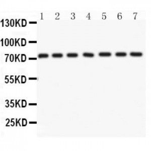
Anti- Lamin A antibody, ASA-B1147, Western blotting
All lanes: Anti Lamin A (ASA-B1147) at 0.5ug/ml
Lane 1: Human Placenta Tissue Lysate at 50ug
Lane 2: SKOV Whole Cell Lysate at 40ug
Lane 3: SW620 Whole Cell Lysate at 40ug
Lane 4: COLO320 Whole Cell Lysate at 40ug
Lane 5: HELA Whole Cell Lysate at 40ug
Lane 6: 293T Whole Cell Lysate at 40ug
Lane 7: A549 Whole Cell Lysate at 40ug
Predicted bind size: 74KD
Observed bind size: 74KD

Anti- Lamin A antibody, ASA-B1147, IHC(P)
IHC(P): Mouse Intestine Tissue
IHC(P): Mouse Intestine Tissue

Anti- Lamin A antibody, ASA-B1147, IHC(P)
IHC(P): Human Mammary Cancer Tissue
IHC(P): Human Mammary Cancer Tissue


