More info
Overview
Long Name | Antibody Type | Antibody Isotype | Host | Species Reactivity | Validated Applications | Purification |
| fibroblast activation protein, alpha | Polyclonal | IgG | Rabbit | Human, Mouse, Rat | IHC-P, IHC-F, WB | Immunogen affinity purified. |
Immunogen | ||||||
| A synthetic peptide corresponding to a sequence at the N-terminus of human Fibroblast activation protein, alpha(57-73aa FFPNWISGQEYLHQSAD), different from the related rat and mouse sequences by three amino acids. | ||||||
Properties
Form | Lyophilized |
Size | 100 µg/vial |
Contents | Antibody is lyophilized with 5 mg BSA, 0.9 mg NaCl, 0.2 mg Na2HPO4, 0.05 mg Thimerosal and 0.05 mg NaN3. *carrier free antibody available upon request. |
Concentration | Reconstitute with 0.2 mL sterile dH2O (500 µg/ml final concentration). |
Storage | At -20 °C for 12 months, as supplied. Store reconstituted antibody at 2-8 °C for one month. For long-term storage, aliquot and store at -20 °C. Avoid repeated freezing and thawing. |
Additional Information Regarding the Antigen
Gene | FAP |
Protein | Seprase |
Uniprot ID | Q12884 |
Function | Cell surface glycoprotein serine protease that participates in extracellular matrix degradation and involved in many cellular processes including tissue remodeling, fibrosis, wound healing, inflammation and tumor growth. Both plasma membrane and soluble forms exhibit post-proline cleaving endopeptidase activity, with a marked preference for Ala/Ser-Gly-Pro-Ser/Asn/Ala consensus sequences, on substrate such as alpha-2-antiplasmin SERPINF2 and SPRY2 (PubMed:14751930, PubMed:16223769, PubMed:16480718, PubMed:16410248, PubMed:17381073, PubMed:18095711, PubMed:21288888, PubMed:24371721). Degrade also gelatin, heat-denatured type I collagen, but not native collagen type I and IV, vibronectin, tenascin, laminin, fibronectin, fibrin or casein (PubMed:9065413, PubMed:2172980, PubMed:7923219, PubMed:10347120, PubMed:10455171, PubMed:12376466, PubMed:16223769, PubMed:16651416, PubMed:18095711). Have also dipeptidyl peptidase activity, exhibiting the ability to hydrolyze the prolyl bond two residues from the N-terminus of synthetic dipeptide substrates provided that the penultimate residue is proline, with a preference for Ala-Pro, Ile-Pro, Gly-Pro, Arg-Pro and Pro-Pro (PubMed:10347120, PubMed:10593948, PubMed:16175601, PubMed:16223769, PubMed:16651416, PubMed:16410248, PubMed:17381073, PubMed:21314817, PubMed:24371721, PubMed:24717288). Natural neuropeptide hormones for dipeptidyl peptidase are the neuropeptide Y (NPY), peptide YY (PYY), substance P (TAC1) and brain natriuretic peptide 32 (NPPB) (PubMed:21314817). The plasma membrane form, in association with either DPP4, PLAUR or integrins, is involved in the pericellular proteolysis of the extracellular matrix (ECM), and hence promotes cell adhesion, migration and invasion through the ECM. Plays a role in tissue remodeling during development and wound healing. Participates in the cell invasiveness towards the ECM in malignant melanoma cancers. Enhances tumor growth progression by increasing angiogenesis, collagen fiber degradation and apoptosis and by reducing antitumor response of the immune system. Promotes glioma cell invasion through the brain parenchyma by degrading the proteoglycan brevican. Acts as a tumor suppressor in melanocytic cells through regulation of cell proliferation and survival in a serine protease activity-independent manner. |
Tissue Specificity | Expressed in adipose tissue. Expressed in the dermal fibroblasts in the fetal skin. Expressed in the granulation tissue of healing wounds and on reactive stromal fibroblast in epithelial cancers. Expressed in activated fibroblast-like synoviocytes from inflamed synovial tissues. Expressed in activated hepatic stellate cells (HSC) and myofibroblasts from cirrhotic liver, but not detected in normal liver. Expressed in glioma cells (at protein level). Expressed in glioblastomas and glioma cells. Isoform 1 and isoform 2 are expressed in melanoma, carcinoma and fibroblast cell lines. |
Sub-cellular localization | Prolyl endopeptidase FAP: Cell surface. |
Sequence Similarities | |
Aliases | 170 kDa melanoma membrane bound gelatinase antibody|170 kDa melanoma membrane-bound gelatinase antibody|DPPIV antibody|FAP antibody|FAPA antibody|Fibroblast activation protein alpha antibody|Integral membrane serine protease antibody|SEPR_HUMAN antibody|Seprase antibody |
Application Details
| Application | Concentration* | Species | Validated Using** |
| Western blot | 0.1-0.5μg/ml | Human, Mouse Rat | AssaySolutio's ECL kit |
| Immunohistochemistry(Paraffin-embedded Section) | 0.5-1μg/ml | Human, Mouse, Rat Rat | AssaySolutio's IHC/ICC Detection kit |
| Immunohistochemistry(Frozen Section) | 0.5-1μg/ml | Rat Human, Mouse | AssaySolutio's IHC/ICC Detection kit |
AssaySolution recommends Rabbit Chemiluminescent WB Detection Kit (AKIT001B) for Western blot, and Rabbit Peroxidase IHC/ICC Detection Kit (AKIT002B) for IHC(P) and IHC(F). *Blocking peptide can be purchased at $65. Contact us for more information

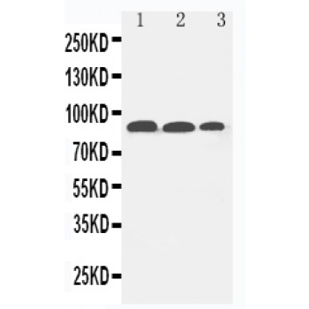
Anti-Fibroblast activation protein, alpha antibody, ASA-B0712, Western blotting
Lane 1: HELA Cell Lysate
Lane 2: SKOV Cell Lysate
Lane 3: NIH3T3 Cell Lysate
Lane 1: HELA Cell Lysate
Lane 2: SKOV Cell Lysate
Lane 3: NIH3T3 Cell Lysate

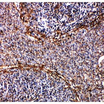
Anti-Fibroblast activation protein, alpha antibody, ASA-B0712, IHC(P)
IHC(P): Rat Spleen Tissue
IHC(P): Rat Spleen Tissue


