More info
Overview
Long Name | Antibody Type | Antibody Isotype | Host | Species Reactivity | Validated Applications | Purification |
| cyclin-dependent kinase 2 | Polyclonal | IgG | Rabbit | Human, Mouse, Rat | IHC-P, WB | Immunogen affinity purified. |
Immunogen | ||||||
| E.coli-derived human Cdk2 recombinant protein (Position: E81-L298). Human Cdk2 shares 98.6% amino acid (aa) sequence identity with rat Cdk2. | ||||||
Properties
Form | Lyophilized |
Size | 100 µg/vial |
Contents | Antibody is lyophilized with 5 mg BSA, 0.9 mg NaCl, 0.2 mg Na2HPO4, 0.05 mg NaN3. *carrier free antibody available upon request. |
Concentration | Reconstitute with 0.2 mL sterile dH2O (500 µg/ml final concentration). |
Storage | At -20 °C for 12 months, as supplied. Store reconstituted antibody at 2-8 °C for one month. For long-term storage, aliquot and store at -20 °C. Avoid repeated freezing and thawing. |
Additional Information Regarding the Antigen
Gene | CDK2 |
Protein | Cyclin-dependent kinase 2 |
Uniprot ID | P24941 |
Function | Serine/threonine-protein kinase involved in the control of the cell cycle; essential for meiosis, but dispensable for mitosis. Phosphorylates CTNNB1, USP37, p53/TP53, NPM1, CDK7, RB1, BRCA2, MYC, NPAT, EZH2. Interacts with cyclins A, B1, B3, D, or E. Triggers duplication of centrosomes and DNA. Acts at the G1-S transition to promote the E2F transcriptional program and the initiation of DNA synthesis, and modulates G2 progression; controls the timing of entry into mitosis/meiosis by controlling the subsequent activation of cyclin B/CDK1 by phosphorylation, and coordinates the activation of cyclin B/CDK1 at the centrosome and in the nucleus. Crucial role in orchestrating a fine balance between cellular proliferation, cell death, and DNA repair in human embryonic stem cells (hESCs). Activity of CDK2 is maximal during S phase and G2; activated by interaction with cyclin E during the early stages of DNA synthesis to permit G1-S transition, and subsequently activated by cyclin A2 (cyclin A1 in germ cells) during the late stages of DNA replication to drive the transition from S phase to mitosis, the G2 phase. EZH2 phosphorylation promotes H3K27me3 maintenance and epigenetic gene silencing. Phosphorylates CABLES1 (By similarity). Cyclin E/CDK2 prevents oxidative stress-mediated Ras-induced senescence by phosphorylating MYC. Involved in G1-S phase DNA damage checkpoint that prevents cells with damaged DNA from initiating mitosis; regulates homologous recombination-dependent repair by phosphorylating BRCA2, this phosphorylation is low in S phase when recombination is active, but increases as cells progress towards mitosis. In response to DNA damage, double-strand break repair by homologous recombination a reduction of CDK2-mediated BRCA2 phosphorylation. Phosphorylation of RB1 disturbs its interaction with E2F1. NPM1 phosphorylation by cyclin E/CDK2 promotes its dissociates from unduplicated centrosomes, thus initiating centrosome duplication. Cyclin E/CDK2-mediated phosphorylation of NPAT at G1-S transition and until prophase stimulates the NPAT- mediated activation of histone gene transcription during S phase. Required for vitamin D-mediated growth inhibition by being itself inactivated. Involved in the nitric oxide- (NO) mediated signaling in a nitrosylation/activation-dependent manner. USP37 is activated by phosphorylation and thus triggers G1-S transition. CTNNB1 phosphorylation regulates insulin internalization. Phosphorylates FOXP3 and negatively regulates its transcriptional activity and protein stability (By similarity). |
Tissue Specificity | |
Sub-cellular localization | Cytoplasm, cytoskeleton, microtubule organizing center, centrosome. Nucleus, Cajal body. Cytoplasm. Endosome. Note: Localized at the centrosomes in late G2 phase after separation of the centrosomes but before the start of prophase. Nuclear-cytoplasmic trafficking is mediated during the inhibition by 1,25-(OH)(2)D(3). |
Sequence Similarities | Belongs to the protein kinase superfamily. CMGC Ser/Thr protein kinase family. CDC2/CDKX subfamily. |
Aliases | Cdc2 related protein kinase antibody|cdc2-related protein kinase antibody|Cdk 2 antibody|CDK2 antibody|CDK2_HUMAN antibody|Cell devision kinase 2 antibody| Cell division kinase 2 antibody|Cell division protein kinase 2 antibody|Cyclin dependent kinase 2 antibody|cyclin dependent kinase 2-alpha antibody|Cyclin-dependent kinase 2 antibody|p33 protein kinase antibody|p33(CDK2) antibody |
Application Details
| Application | Concentration* | Species | Validated Using** |
| Western blot | 0.1-0.5μg/ml | Human, Rat | AssaySolutio's ECL kit |
| Immunohistochemistry(Paraffin-embedded Section) | 0.5-1μg/ml | Human, Mouse, Rat | AssaySolutio's IHC/ICC Detection kit |
AssaySolution recommends Rabbit Chemiluminescent WB Detection Kit (AKIT001B) for Western blot, and Rabbit Peroxidase IHC/ICC Detection Kit (AKIT002B) for IHC(P). *Blocking peptide can be purchased at $65. Contact us for more information

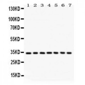
Anti- Cdk2 antibody, ASA-B0430, Western blotting
All lanes: Anti Cdk2 (ASA-B0430) at 0.5ug/ml
Lane 1: Rat Kidney Tissue Lysate at 50ug
Lane 2: Rat Liver Tissue Lysate at 50ug
Lane 3: Human Placenta Tissue Lysate at 50ug
Lane 4: HELA Whole Cell Lysate at 40ug
Lane 5: HUT Whole Cell Lysate at 40ug
Lane 6: JURKAT Whole Cell Lysate at 40ug
Lane 7: A549 Whole Cell Lysate at 40ug
Predicted bind size: 34KD
Observed bind size: 34KD

Anti- Cdk2 antibody, ASA-B0430,IHC(P)
IHC(P): Mouse Testis Tissue
IHC(P): Mouse Testis Tissue

Anti- Cdk2 antibody, ASA-B0430,IHC(P)
IHC(P): Rat Testis Tissue
IHC(P): Rat Testis Tissue


