More info
Overview
Long Name | Antibody Type | Antibody Isotype | Host | Species Reactivity | Validated Applications | Purification |
| CD63 molecule | Polyclonal | IgG | Rabbit | Human | IHC-P, WB | Immunogen affinity purified. |
Immunogen | ||||||
| E.coli-derived human CD63 recombinant protein (Position: E97-M238). Human CD63 shares 74% and 73% amino acid (aa) sequence identity with mouse and rat CD63, respectively. | ||||||
Properties
Form | Lyophilized |
Size | 100 µg/vial |
Contents | Antibody is lyophilized with 5 mg BSA, 0.9 mg NaCl, 0.2 mg Na2HPO4, 0.05 mg NaN3. *carrier free antibody available upon request. |
Concentration | Reconstitute with 0.2 mL sterile dH2O (500 µg/ml final concentration). |
Storage | At -20 °C for 12 months, as supplied. Store reconstituted antibody at 2-8 °C for one month. For long-term storage, aliquot and store at -20 °C. Avoid repeated freezing and thawing. |
Additional Information Regarding the Antigen
Gene | CD63 |
Protein | CD63 antigen |
Uniprot ID | P08962 |
Function | Functions as cell surface receptor for TIMP1 and plays a role in the activation of cellular signaling cascades. Plays a role in the activation of ITGB1 and integrin signaling, leading to the activation of AKT, FAK/PTK2 and MAP kinases. Promotes cell survival, reorganization of the actin cytoskeleton, cell adhesion, spreading and migration, via its role in the activation of AKT and FAK/PTK2. Plays a role in VEGFA signaling via its role in regulating the internalization of KDR/VEGFR2. Plays a role in intracellular vesicular transport processes, and is required for normal trafficking of the PMEL luminal domain that is essential for the development and maturation of melanocytes. Plays a role in the adhesion of leukocytes onto endothelial cells via its role in the regulation of SELP trafficking. May play a role in mast cell degranulation in response to Ms4a2/FceRI stimulation, but not in mast cell degranulation in response to other stimuli. |
Tissue Specificity | Detected in platelets (at protein level). Dysplastic nevi, radial growth phase primary melanomas, hematopoietic cells, tissue macrophages. |
Sub-cellular localization | Cell membrane; Multi-pass membrane protein. Lysosome membrane; Multi-pass membrane protein. Late endosome membrane; Multi-pass membrane protein. Endosome, multivesicular body. Melanosome. Note: Also found in Weibel-Palade bodies of endothelial cells. Located in platelet dense granules. Detected in a subset of pre-melanosomes. Detected on intralumenal vesicles (ILVs) within multivesicular bodies. |
Sequence Similarities | |
Aliases | Lysosomal associated membrane protein 3 antibody|CD 63 antibody|CD63 antibody|CD63 antigen (melanoma 1 antigen) antibody|CD63 antigen antibody|CD63 antigen melanoma 1 antigen antibody|CD63 molecule antibody|CD63_HUMAN antibody|gp55 antibody|granulophysin antibody|LAMP 3 antibody|LAMP-3 antibody|LAMP3 antibody|LIMP antibody|Lysosomal-associated membrane protein 3 antibody|lysosome associated membrane glycoprotein 3 antibody|Mast cell antigen AD1 antibody|ME491 antibody|melanoma 1 antigen antibody|Melanoma associated antigen ME491 antibody|Melanoma associated antigen MLA1 antibody|Melanoma-associated antigen ME491 antibody|MGC72893 antibody|MLA 1 antibody|MLA1 antibody|NGA antibody|ocular melanoma associated antigen antibody|Ocular melanoma-associated antigen antibody|OMA81H antibody|PTLGP40 antibody|Tetraspanin 30 antibody|Tetraspanin-30 antibody|Tspan 30 antibody|Tspan-30 antibody|TSPAN30 antibody |
Application Details
| Application | Concentration* | Species | Validated Using** |
| Western blot | 0.1-0.5μg/ml | Human | AssaySolutio's ECL kit |
| Immunohistochemistry(Paraffin-embedded Section) | 0.5-1μg/ml | Human | AssaySolutio's IHC/ICC Detection kit |
AssaySolution recommends Rabbit Chemiluminescent WB Detection Kit (AKIT001B) for Western blot, and Rabbit Peroxidase IHC/ICC Detection Kit (AKIT002B) for IHC(P). *Blocking peptide can be purchased at $65. Contact us for more information

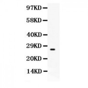
Anti- CD63 antibody, ASA-B0397, Western blotting
All lanes: Anti CD63 (ASA-B0397) at 0.5ug/ml
WB: Recombinant Human CD63 Protein 0.5ng
Predicted bind size: 26KD
Observed bind size: 26KD
All lanes: Anti CD63 (ASA-B0397) at 0.5ug/ml
WB: Recombinant Human CD63 Protein 0.5ng
Predicted bind size: 26KD
Observed bind size: 26KD

Anti- CD63 antibody, ASA-B0397, Western blotting
All lanes: Anti CD63 (ASA-B0397) at 0.5ug/ml
WB: HELA Whole Cell Lysate at 40ug
Predicted bind size: 26KD
Observed bind size: 35KD
All lanes: Anti CD63 (ASA-B0397) at 0.5ug/ml
WB: HELA Whole Cell Lysate at 40ug
Predicted bind size: 26KD
Observed bind size: 35KD

Anti- CD63 antibody, ASA-B0397, IHC(P)
IHC(P): Human Intestinal Cancer Tissue
IHC(P): Human Intestinal Cancer Tissue


