More info
Overview
Long Name | Antibody Type | Antibody Isotype | Host | Species Reactivity | Validated Applications | Purification |
| TEK tyrosine kinase, endothelial | Polyclonal | IgG | Rabbit | Human | WB | Immunogen affinity purified. |
Immunogen | ||||||
| E.coli-derived human TIE2 recombinant protein (Position: Q641-I830). Human TIE2 shares 91% amino acid (aa) sequence identity with mouse TIE2. | ||||||
Properties
Form | Lyophilized |
Size | 100 µg/vial |
Contents | Antibody is lyophilized with 5 mg BSA, 0.9 mg NaCl, 0.2 mg Na2HPO4, 0.05 mg NaN3. *carrier free antibody available upon request. |
Concentration | Reconstitute with 0.2 mL sterile dH2O (500 µg/ml final concentration). |
Storage | At -20 °C for 12 months, as supplied. Store reconstituted antibody at 2-8 °C for one month. For long-term storage, aliquot and store at -20 °C. Avoid repeated freezing and thawing. |
Additional Information Regarding the Antigen
Gene | TEK |
Protein | Angiopoietin-1 receptor |
Uniprot ID | Q02763 |
Function | Tyrosine-protein kinase that acts as cell-surface receptor for ANGPT1, ANGPT2 and ANGPT4 and regulates angiogenesis, endothelial cell survival, proliferation, migration, adhesion and cell spreading, reorganization of the actin cytoskeleton, but also maintenance of vascular quiescence. Has anti-inflammatory effects by preventing the leakage of proinflammatory plasma proteins and leukocytes from blood vessels. Required for normal angiogenesis and heart development during embryogenesis. Required for post- natal hematopoiesis. After birth, activates or inhibits angiogenesis, depending on the context. Inhibits angiogenesis and promotes vascular stability in quiescent vessels, where endothelial cells have tight contacts. In quiescent vessels, ANGPT1 oligomers recruit TEK to cell-cell contacts, forming complexes with TEK molecules from adjoining cells, and this leads to preferential activation of phosphatidylinositol 3-kinase and the AKT1 signaling cascades. In migrating endothelial cells that lack cell-cell adhesions, ANGT1 recruits TEK to contacts with the extracellular matrix, leading to the formation of focal adhesion complexes, activation of PTK2/FAK and of the downstream kinases MAPK1/ERK2 and MAPK3/ERK1, and ultimately to the stimulation of sprouting angiogenesis. ANGPT1 signaling triggers receptor dimerization and autophosphorylation at specific tyrosine residues that then serve as binding sites for scaffold proteins and effectors. Signaling is modulated by ANGPT2 that has lower affinity for TEK, can promote TEK autophosphorylation in the absence of ANGPT1, but inhibits ANGPT1-mediated signaling by competing for the same binding site. Signaling is also modulated by formation of heterodimers with TIE1, and by proteolytic processing that gives rise to a soluble TEK extracellular domain. The soluble extracellular domain modulates signaling by functioning as decoy receptor for angiopoietins. TEK phosphorylates DOK2, GRB7, GRB14, PIK3R1; SHC1 and TIE1. |
Tissue Specificity | Detected in umbilical vein endothelial cells. Proteolytic processing gives rise to a soluble extracellular domain that is detected in blood plasma (at protein level). Predominantly expressed in endothelial cells and their progenitors, the angioblasts. Has been directly found in placenta and lung, with a lower level in umbilical vein endothelial cells, brain and kidney. |
Sub-cellular localization | Cell membrane; Single-pass type I membrane protein. Cell junction. Cell junction, focal adhesion. Cytoplasm, cytoskeleton. Secreted. Note: Recruited to cell-cell contacts in quiescent endothelial cells. Colocalizes with the actin cytoskeleton and at actin stress fibers during cell spreading. Recruited to the lower surface of migrating cells, especially the rear end of the cell. Proteolytic processing gives rise to a soluble extracellular domain that is secreted. |
Sequence Similarities | Belongs to the protein kinase superfamily. Tyr protein kinase family. Tie subfamily. |
Aliases | Angiopoietin 1 receptor antibody|Angiopoietin-1 receptor antibody|CD202b antibody|CD202b antigen antibody|CD202b antigen antibody|Endothelial tyrosine kinase antibody|Endothelium specific receptor tyrosine kinase 2 antibody|hTIE 2 antibody|hTIE 2 antibody|hTIE2 antibody|hTIE2 antibody|Hyk antibody|p140 TEK antibody|p140 TEK antibody|Soluble TIE2 variant 1 antibody|Soluble TIE2 variant 2 antibody|TEK antibody|tek tyrosine kinase antibody|TEK tyrosine kinase endothelial antibody|tek tyrosine kinase, endothelial antibody|TIE 2 antibody|TIE2_HUMAN antibody|Tunica interna endothelial cell kinase antibody|Tyrosine kinase with Ig and EGF homology domains 2 antibody|Tyrosine protein kinase receptor TEK antibody|Tyrosine protein kinase receptor TIE 2 antibody|Tyrosine protein kinase receptor TIE 2 antibody|Tyrosine-protein kinase receptor TEK antibody|Tyrosine-protein kinase receptor TIE-2 antibody|Venous malformations multiple cutaneous and mucosal antibody|VMCM 1 antibody|VMCM antibody|VMCM1 antibody |
Application Details
| Application | Concentration* | Species | Validated Using** |
| Western blot | 0.1-0.5μg/ml | Human | AssaySolutio's ECL kit |
AssaySolution recommends Rabbit Chemiluminescent WB Detection Kit (AKIT001B) for Western blot. *Blocking peptide can be purchased at $65. Contact us for more information
Anti-TIE2 antibody, ASA-B1859--1.jpg
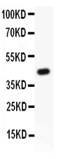
All lanes: Anti TIE2 (ASA-B1859) at 0.5ug/ml
WB: Recombinant Human TIE2 Protein 0.5ng
Predicted bind size: 47KD
Observed bind size: 47KD
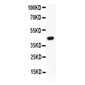
All lanes: Anti TIE2 (ASA-B1859) at 0.5ug/ml
WB: Recombinant Human TIE2 Protein 0.5ng
Predicted bind size: 47KD
Observed bind size: 47KD
Anti-TIE2 antibody, ASA-B1859--2.jpg
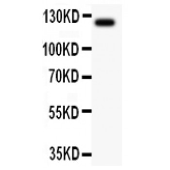
All lanes: Anti TIE2 (ASA-B1859) at 0.5ug/ml
WB: MCF-7 Whole Cell Lysate at 40ug
Predicted bind size: 125KD
Observed bind size: 125KD
All lanes: Anti TIE2 (ASA-B1859) at 0.5ug/ml
WB: MCF-7 Whole Cell Lysate at 40ug
Predicted bind size: 125KD
Observed bind size: 125KD