More info
Overview
Long Name | Antibody Type | Antibody Isotype | Host | Species Reactivity | Validated Applications | Purification |
| podoplanin | Polyclonal | IgG | Rabbit | Human | WB | Immunogen affinity purified. |
Immunogen | ||||||
| A synthetic peptide corresponding to a sequence at the C-terminus of human Podoplanin/gp36(227-238aa VVMRKMSGRYSP). | ||||||
Properties
Form | Lyophilized |
Size | 100 µg/vial |
Contents | Antibody is lyophilized with 5 mg BSA, 0.9 mg NaCl, 0.2 mg Na2HPO4, 0.05 mg Thimerosal and 0.05 mg NaN3. *carrier free antibody available upon request. |
Concentration | Reconstitute with 0.2 mL sterile dH2O (500 µg/ml final concentration). |
Storage | At -20 °C for 12 months, as supplied. Store reconstituted antibody at 2-8 °C for one month. For long-term storage, aliquot and store at -20 °C. Avoid repeated freezing and thawing. |
Additional Information Regarding the Antigen
Gene | PDPN |
Protein | Podoplanin |
Uniprot ID | Q86YL7 |
Function | May be involved in cell migration and/or actin cytoskeleton organization. When expressed in keratinocytes, induces changes in cell morphology with transfected cells showing an elongated shape, numerous membrane protrusions, major reorganization of the actin cytoskeleton, increased motility and decreased cell adhesion. Required for normal lung cell proliferation and alveolus formation at birth. Induces platelet aggregation. Does not have any effect on folic acid or amino acid transport. Does not function as a water channel or as a regulator of aquaporin-type water channels. |
Tissue Specificity | Highly expressed in placenta, lung, skeletal muscle and brain. Weakly expressed in brain, kidney and liver. In placenta, expressed on the apical plasma membrane of endothelium. In lung, expressed in alveolar epithelium. Up-regulated in colorectal tumors and expressed in 25% of early oral squamous cell carcinomas. |
Sub-cellular localization | Membrane ; Single-pass type I membrane protein . Cell projection, filopodium membrane ; Single-pass type I membrane protein . Cell projection, lamellipodium membrane ; Single-pass type I membrane protein . Cell projection, microvillus membrane ; Single-pass type I membrane protein . Cell projection, ruffle membrane ; Single-pass type I membrane protein . Note: Localized to actin-rich microvilli and plasma membrane projections such as filopodia, lamellipodia and ruffles. |
Sequence Similarities | Belongs to the podoplanin family. |
Aliases | AGGRUS antibody|GLYCOPROTEIN 36 KD antibody|Glycoprotein 36 antibody|gp 36 antibody|GP 38 antibody|GP 40 antibody|gp36 antibody|GP38 antibody|GP40 antibody|HT1A 1 antibody|HT1A1 antibody|hT1alpha1 antibody|hT1alpha2 antibody|Lung type I cell membrane associated glycoprotein antibody|Lung type I cell membrane associated glycoprotein isoform a antibody|Lung type I cell membrane associated glycoprotein T1A 2 antibody|OTS 8 antibody|OTS8 antibody|OTTHUMP00000009640 antibody|OTTHUMP00000044504 antibody|PA2.26 antibody|PA2.26 antigen antibody|PDPN antibody|PDPN_HUMAN antibody|Podoplanin antibody|PSEC0003 antibody|PSEC0025 antibody|T1 alpha antibody|T1 ALPHA GENE antibody|T1-alpha antibody|T1A 2 antibody|T1A antibody|TIA 2 antibody|TIA2 antibody |
Application Details
| Application | Concentration* | Species | Validated Using** |
| Western blot | 0.1-0.5μg/ml | Human | AssaySolutio's ECL kit |
AssaySolution recommends Rabbit Chemiluminescent WB Detection Kit (AKIT001B) for Western blot. *Blocking peptide can be purchased at $65. Contact us for more information
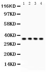
Anti-Podoplanin/gp36 antibody, ASA-B1552, Western blotting
All lanes: Anti PDPN (ASA-B1552) at 0.5ug/ml
Lane 1: A549 Whole Cell Lysate at 40ug
Lane 2: SMMC Whole Cell Lysate at 40ug
Lane 3: 293T Whole Cell Lysate at 40ug
Lane 4: HELA Whole Cell Lysate at 40ug
Predicted bind size: 36KD
Observed bind size: 36KD
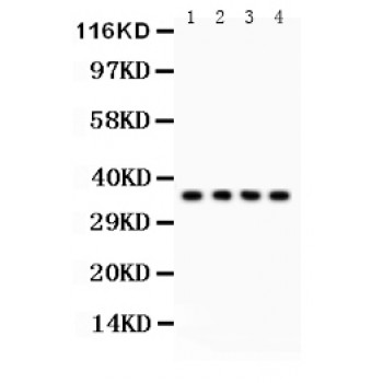
All lanes: Anti PDPN (ASA-B1552) at 0.5ug/ml
Lane 1: A549 Whole Cell Lysate at 40ug
Lane 2: SMMC Whole Cell Lysate at 40ug
Lane 3: 293T Whole Cell Lysate at 40ug
Lane 4: HELA Whole Cell Lysate at 40ug
Predicted bind size: 36KD
Observed bind size: 36KD