More info
Overview
Long Name | Antibody Type | Antibody Isotype | Host | Species Reactivity | Validated Applications | Purification |
| MDM2 oncogene, E3 ubiquitin protein ligase | Polyclonal | IgG | Rabbit | Human, Mouse, Rat | WB | Immunogen affinity purified. |
Immunogen | ||||||
| A synthetic peptide corresponding to a sequence at the C-terminus of human MDM2(343-358aa EKAKLENSTQAEEGFD), different from the related mouse sequence by two amino acids, and from the related rat sequence by three amino acids. | ||||||
Properties
Form | Lyophilized |
Size | 100 µg/vial |
Contents | Antibody is lyophilized with 5 mg BSA, 0.9 mg NaCl, 0.2 mg Na2HPO4, 0.05 mg Thimerosal and 0.05 mg NaN3. *carrier free antibody available upon request. |
Concentration | Reconstitute with 0.2 mL sterile dH2O (500 µg/ml final concentration). |
Storage | At -20 °C for 12 months, as supplied. Store reconstituted antibody at 2-8 °C for one month. For long-term storage, aliquot and store at -20 °C. Avoid repeated freezing and thawing. |
Additional Information Regarding the Antigen
Gene | MDM2 |
Protein | E3 ubiquitin-protein ligase Mdm2 |
Uniprot ID | Q00987 |
Function | E3 ubiquitin-protein ligase that mediates ubiquitination of p53/TP53, leading to its degradation by the proteasome. Inhibits p53/TP53- and p73/TP73-mediated cell cycle arrest and apoptosis by binding its transcriptional activation domain. Also acts as a ubiquitin ligase E3 toward itself and ARRB1. Permits the nuclear export of p53/TP53. Promotes proteasome-dependent ubiquitin-independent degradation of retinoblastoma RB1 protein. Inhibits DAXX-mediated apoptosis by inducing its ubiquitination and degradation. Component of the TRIM28/KAP1-MDM2-p53/TP53 complex involved in stabilizing p53/TP53. Also component of the TRIM28/KAP1-ERBB4-MDM2 complex which links growth factor and DNA damage response pathways. Mediates ubiquitination and subsequent proteasome degradation of DYRK2 in nucleus. Ubiquitinates IGF1R and SNAI1 and promotes them to proteasomal degradation. |
Tissue Specificity | Ubiquitous. Isoform Mdm2-A, isoform Mdm2-B, isoform Mdm2-C, isoform Mdm2-D, isoform Mdm2-E, isoform Mdm2-F and isoform Mdm2-G are observed in a range of cancers but absent in normal tissues. |
Sub-cellular localization | Nucleus, nucleoplasm. Cytoplasm. Nucleus, nucleolus. Note: Expressed predominantly in the nucleoplasm. Interaction with ARF(P14) results in the localization of both proteins to the nucleolus. The nucleolar localization signals in both ARF(P14) and MDM2 may be necessary to allow efficient nucleolar localization of both proteins. Colocalizes with RASSF1 isoform A in the nucleus. |
Sequence Similarities | Belongs to the MDM2/MDM4 family. |
Aliases | Double minute 2 protein antibody|E3 ubiquitin-protein ligase Mdm2 antibody|Hdm 2 antibody|HDM2 antibody|MDM 2 antibody|MDM2 antibody|Mdm2 transformed 3T3 cell double minute 2 p53 binding protein(mouse) binding protein 104kDa antibody|MDM2_HUMAN antibody|MDM2BP antibody|Mouse Double Minute 2 antibody|MTBP antibody|Murine Double Minute Chromosome 2 antibody|Oncoprotein Mdm2 antibody|p53 Binding Protein Mdm2 antibody|p53-binding protein Mdm2 antibody|Ubiquitin protein ligase E3 Mdm2 antibody |
Application Details
| Application | Concentration* | Species | Validated Using** |
| Western blot | 0.1-0.5μg/ml | Human, Mouse, Rat | AssaySolutio's ECL kit |
AssaySolution recommends Rabbit Chemiluminescent WB Detection Kit (AKIT001B) for Western blot. *Blocking peptide can be purchased at $65. Contact us for more information
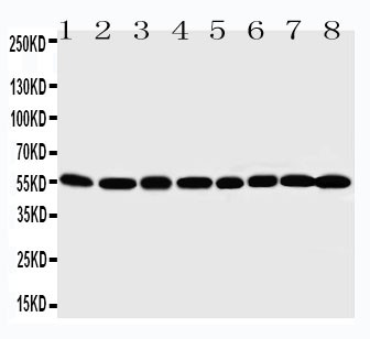
Anti-MDM2 antibody, ASA-B1247, Western blotting
Lane 1: Rat Testis Tissue Lysate
Lane 2: Rat Brain Tissue Lysate
Lane 3: Rat Heart Tissue Lysate
Lane 4: SKOV-3 Cell Lysate
Lane 5: CLOL320 Cell Lysate
Lane 6: HELA Cell Lysate
Lane 7: HEPA Cell Lysate
Lane 8: COS7 Cell Lysate
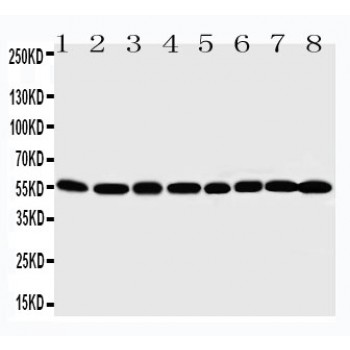
Lane 1: Rat Testis Tissue Lysate
Lane 2: Rat Brain Tissue Lysate
Lane 3: Rat Heart Tissue Lysate
Lane 4: SKOV-3 Cell Lysate
Lane 5: CLOL320 Cell Lysate
Lane 6: HELA Cell Lysate
Lane 7: HEPA Cell Lysate
Lane 8: COS7 Cell Lysate