More info
Overview
Long Name | Antibody Type | Antibody Isotype | Host | Species Reactivity | Validated Applications | Purification |
| neural precursor cell expressed, developmentally down-regulated 9 | Polyclonal | IgG | Rabbit | Human | WB | Immunogen affinity purified. |
Immunogen | ||||||
| E.coli-derived human HEF1 recombinant protein (Position: K273-E421). Human HEF1 shares 83% amino acid (aa) sequence identity with mouse HEF1. | ||||||
Properties
Form | Lyophilized |
Size | 100 µg/vial |
Contents | Antibody is lyophilized with 5 mg BSA, 0.9 mg NaCl, 0.2 mg Na2HPO4, 0.05 mg NaN3. *carrier free antibody available upon request. |
Concentration | Reconstitute with 0.2 mL sterile dH2O (500 µg/ml final concentration). |
Storage | At -20 °C for 12 months, as supplied. Store reconstituted antibody at 2-8 °C for one month. For long-term storage, aliquot and store at -20 °C. Avoid repeated freezing and thawing. |
Additional Information Regarding the Antigen
Gene | NEDD9 |
Protein | Enhancer of filamentation 1 |
Uniprot ID | Q14511 |
Function | Docking protein which plays a central coordinating role for tyrosine-kinase-based signaling related to cell adhesion. May function in transmitting growth control signals between focal adhesions at the cell periphery and the mitotic spindle in response to adhesion or growth factor signals initiating cell proliferation. May play an important role in integrin beta-1 or B cell antigen receptor (BCR) mediated signaling in B- and T-cells. Integrin beta-1 stimulation leads to recruitment of various proteins including CRK, NCK and SHPTP2 to the tyrosine phosphorylated form. |
Tissue Specificity | Widely expressed. Higher levels detected in kidney, lung, and placenta. Also detected in T-cells, B-cells and diverse cell lines. The protein has been detected in lymphocytes, in diverse cell lines, and in lung tissues. |
Sub-cellular localization | Cytoplasm, cell cortex. Nucleus. Golgi apparatus. Cell projection, lamellipodium. Cytoplasm. Cell junction, focal adhesion. Note: Localizes to both the cell nucleus and the cell periphery and is differently localized in fibroblasts and epithelial cells. In fibroblasts is predominantly nuclear and in some cells is present in the Golgi apparatus. In epithelial cells localized predominantly in the cell periphery with particular concentration in lamellipodia but is also found in the nucleus. Isoforms p105 and p115 are predominantly cytoplasmic and associate with focal adhesions while p55 associates with mitotic spindle. |
Sequence Similarities | Belongs to the CAS family. |
Aliases | CAS L antibody|Cas like docking antibody|Cas scaffolding protein family member 2 antibody|CAS-L antibody|CAS2 antibody|CASL antibody|CASL_HUMAN antibody|CASS2 antibody|Crk associated substrate related antibody|Crk associated substrate related protein antibody|CRK-associated substrate-related protein antibody|dJ49G10.2 (Enhancer of Filamentation 1 (HEF1)) antibody|dJ49G10.2 antibody|dJ761I2.1 (enhancer of filamentation (HEF1)) antibody|dJ761I2.1 antibody|Enhancer of filamentation 1 antibody|Enhancer of filamentation 1 p55 antibody|HEF 1 antibody|HEF1 antibody|NEDD 9 antibody|NEDD-9 antibody|NEDD9 antibody|NEDD9 protein antibody|Neural cell expressed developmentally down regulated 9 antibody|Neural precursor cell expressed developmentally down regulated 9 antibody|Neural precursor cell expressed developmentally down-regulated protein 9 antibody|NY REN 12 antigen antibody|p105 antibody|Protein NEDD9 antibody|Renal carcinoma antigen NY-REN-12 antibody |
Application Details
| Application | Concentration* | Species | Validated Using** |
| Western blot | 0.1-0.5μg/ml | Human | AssaySolutio's ECL kit |
AssaySolution recommends Rabbit Chemiluminescent WB Detection Kit (AKIT001B) for Western blot. *Blocking peptide can be purchased at $65. Contact us for more information

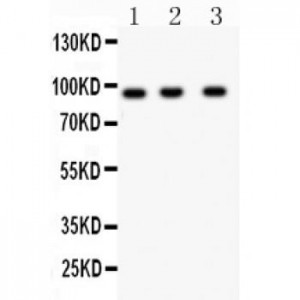
Anti- HEF1 antibody, ASA-B0850, Western blotting
All lanes: Anti HEF1 (ASA-B0850) at 0.5ug/ml
Lane 1: JURKAT Whole Cell Lysate at 40ug
Lane 2: CEM Whole Cell Lysate at 40ug
Lane 3: RAJI Whole Cell Lysate at 40ug
Predicted bind size: 93KD
Observed bind size: 93KD
All lanes: Anti HEF1 (ASA-B0850) at 0.5ug/ml
Lane 1: JURKAT Whole Cell Lysate at 40ug
Lane 2: CEM Whole Cell Lysate at 40ug
Lane 3: RAJI Whole Cell Lysate at 40ug
Predicted bind size: 93KD
Observed bind size: 93KD


