More info
Overview
Long Name | Antibody Type | Antibody Isotype | Host | Species Reactivity | Validated Applications | Purification |
| amyloid beta (A4) precursor protein-binding, family B, member 1 (Fe65) | Polyclonal | IgG | Rabbit | Human, Mouse | WB | Immunogen affinity purified. |
Immunogen | ||||||
| E.coli-derived human FE65 recombinant protein (Position: Q295-A613). Human FE65 shares 95%and 96% amino acid (aa) sequence identity with mouse and rat FE65, respectively. | ||||||
Properties
Form | Lyophilized |
Size | 100 µg/vial |
Contents | Antibody is lyophilized with 5 mg BSA, 0.9 mg NaCl, 0.2 mg Na2HPO4, 0.05 mg NaN3. *carrier free antibody available upon request. |
Concentration | Reconstitute with 0.2 mL sterile dH2O (500 µg/ml final concentration). |
Storage | At -20 °C for 12 months, as supplied. Store reconstituted antibody at 2-8 °C for one month. For long-term storage, aliquot and store at -20 °C. Avoid repeated freezing and thawing. |
Additional Information Regarding the Antigen
Gene | APBB1 |
Protein | Amyloid beta A4 precursor protein-binding family B member 1 |
Uniprot ID | O00213 |
Function | Transcription coregulator that can have both coactivator and corepressor functions. Adapter protein that forms a transcriptionally active complex with the gamma-secretase-derived amyloid precursor protein (APP) intracellular domain. Plays a central role in the response to DNA damage by translocating to the nucleus and inducing apoptosis. May act by specifically recognizing and binding histone H2AX phosphorylated on 'Tyr-142' (H2AXY142ph) at double-strand breaks (DSBs), recruiting other pro- apoptosis factors such as MAPK8/JNK1. Required for histone H4 acetylation at double-strand breaks (DSBs). Its ability to specifically bind modified histones and chromatin modifying enzymes such as KAT5/TIP60, probably explains its trancription activation activity. Function in association with TSHZ3, SET and HDAC factors as a transcriptional repressor, that inhibits the expression of CASP4. Associates with chromatin in a region surrounding the CASP4 transcriptional start site(s). |
Tissue Specificity | Highly expressed in brain; strongly reduced in post-mortem elderly subjects with Alzheimer disease. |
Sub-cellular localization | Cell membrane. Cytoplasm. Nucleus. Cell projection, growth cone . Nucleus speckle. Note: Colocalizes with TSHZ3 in axonal growth cone (By similarity). In normal conditions, it mainly localizes to the cytoplasm, while a small fraction is tethered to the cell membrane via its interaction with APP. Following exposure to DNA damaging agents, it is released from cell membrane and translocates to the nucleus. Nuclear translocation is under the regulation of APP. Colocalizes with TSHZ3 in the nucleus. Colocalizes with NEK6 at the nuclear speckles. Phosphorylation at Ser-610 by SGK1 promotes its localization to the nucleus (By similarity). |
Sequence Similarities | Contains 2 PID domains. |
Aliases | Adaptor protein FE65a2 antibody|Amyloid beta (A4) precursor protein binding family B member 1 antibody|Amyloid Beta A4 Precursor Protein Binding Family B antibody|Amyloid beta A4 precursor protein binding family B member 1 antibody|Amyloid beta A4 precursor protein-binding family B member 1 antibody|Amyloid beta precursor protein binding family B member 1 antibody|APBB 1 antibody|APBB1 antibody|APBB1_HUMAN antibody|FE 65 antibody|Fe65 protein antibody|Protein Fe65 antibody|RIR antibody|stat like protein antibody |
Application Details
| Application | Concentration* | Species | Validated Using** |
| Western blot | 0.1-0.5μg/ml | Human, Mouse | AssaySolutio's ECL kit |
AssaySolution recommends Rabbit Chemiluminescent WB Detection Kit (AKIT001B) for Western blot. *Blocking peptide can be purchased at $65. Contact us for more information

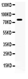
Anti- FE65 antibody, ASA-B0693, Western blotting
All lanes: Anti (ASA-B0693) at 0.5ug/ml
WB: Mouse Brain Tissue Lysate at 50ug
Predicted bind size: 77KD
Observed bind size: 77KD
All lanes: Anti (ASA-B0693) at 0.5ug/ml
WB: Mouse Brain Tissue Lysate at 50ug
Predicted bind size: 77KD
Observed bind size: 77KD