

More info
Overview
Long Name | Antibody Type | Antibody Isotype | Host | Species Reactivity | Validated Applications | Purification |
| v-erb-a erythroblastic leukemia viral oncogene homolog 4(avian) | Polyclonal | IgG | Rabbit | Human, Mouse, Rat | WB | Immunogen affinity purified. |
Immunogen | ||||||
| A synthetic peptide corresponding to a sequence at the N-terminus of human ErbB 4(35-51aa KLSSLSDLEQQYRALRK), identical to the related rat and mouse sequences. | ||||||
Properties
Form | Lyophilized |
Size | 100 µg/vial |
Contents | Antibody is lyophilized with 5 mg BSA, 0.9 mg NaCl, 0.2 mg Na2HPO4, 0.05 mg Thimerosal and 0.05 mg NaN3. *carrier free antibody available upon request. |
Concentration | Reconstitute with 0.2 mL sterile dH2O (500 µg/ml final concentration). |
Storage | At -20 °C for 12 months, as supplied. Store reconstituted antibody at 2-8 °C for one month. For long-term storage, aliquot and store at -20 °C. Avoid repeated freezing and thawing. |
Additional Information Regarding the Antigen
Gene | ERBB4 |
Protein | Receptor tyrosine-protein kinase erbB-4 |
Uniprot ID | Q15303 |
Function | Tyrosine-protein kinase that plays an essential role as cell surface receptor for neuregulins and EGF family members and regulates development of the heart, the central nervous system and the mammary gland, gene transcription, cell proliferation, differentiation, migration and apoptosis. Required for normal cardiac muscle differentiation during embryonic development, and for postnatal cardiomyocyte proliferation. Required for normal development of the embryonic central nervous system, especially for normal neural crest cell migration and normal axon guidance. Required for mammary gland differentiation, induction of milk proteins and lactation. Acts as cell-surface receptor for the neuregulins NRG1, NRG2, NRG3 and NRG4 and the EGF family members BTC, EREG and HBEGF. Ligand binding triggers receptor dimerization and autophosphorylation at specific tyrosine residues that then serve as binding sites for scaffold proteins and effectors. Ligand specificity and signaling is modulated by alternative splicing, proteolytic processing, and by the formation of heterodimers with other ERBB family members, thereby creating multiple combinations of intracellular phosphotyrosines that trigger ligand- and context-specific cellular responses. Mediates phosphorylation of SHC1 and activation of the MAP kinases MAPK1/ERK2 and MAPK3/ERK1. Isoform JM-A CYT-1 and isoform JM-B CYT-1 phosphorylate PIK3R1, leading to the activation of phosphatidylinositol 3-kinase and AKT1 and protect cells against apoptosis. Isoform JM-A CYT-1 and isoform JM-B CYT-1 mediate reorganization of the actin cytoskeleton and promote cell migration in response to NRG1. Isoform JM-A CYT-2 and isoform JM-B CYT-2 lack the phosphotyrosine that mediates interaction with PIK3R1, and hence do not phosphorylate PIK3R1, do not protect cells against apoptosis, and do not promote reorganization of the actin cytoskeleton and cell migration. Proteolytic processing of isoform JM-A CYT-1 and isoform JM-A CYT-2 gives rise to the corresponding soluble intracellular domains (4ICD) that translocate to the nucleus, promote nuclear import of STAT5A, activation of STAT5A, mammary epithelium differentiation, cell proliferation and activation of gene expression. The ERBB4 soluble intracellular domains (4ICD) colocalize with STAT5A at the CSN2 promoter to regulate transcription of milk proteins during lactation. The ERBB4 soluble intracellular domains can also translocate to mitochondria and promote apoptosis. |
Tissue Specificity | Expressed at highest levels in brain, heart, kidney, in addition to skeletal muscle, parathyroid, cerebellum, pituitary, spleen, testis and breast. Lower levels in thymus, lung, salivary gland, and pancreas. Isoform JM-A CYT-1 and isoform JM-B CYT-1 are expressed in cerebellum, but only the isoform JM-B is expressed in the heart. |
Sub-cellular localization | Cell membrane. |
Sequence Similarities | Belongs to the protein kinase superfamily. Tyr protein kinase family. EGF receptor subfamily. |
Aliases | 4ICD antibody|Avian erythroblastic leukemia viral oncogene homolog 4 antibody|Avian erythroblastic leukemia viral v erb b2 oncogene homolog 4 antibody|E4ICD antibody|EC 2.7.10.1 antibody|ErbB4 antibody|ERBB4 intracellular domain antibody|ERBB4_HUMAN antibody|HER 4 antibody|HER4 antibody|Mer4 antibody|MGC138404 antibody|Oncogene ERBB4 antibody|P180erbB4 antibody|Proto-oncogene-like protein c-ErbB-4 antibody|Receptor protein tyrosine kinase erbB 4 precursor antibody|Receptor tyrosine protein kinase erbB 4 antibody|s80HER4 antibody|Tyrosine kinase type cell surface receptor HER4 antibody|Tyrosine kinase-type cell surface receptor HER4 antibody|v erb a avian erythroblastic leukemia viral oncogene homolog like 4 antibody|v erb a erythroblastic leukemia viral oncogene homolog 4 antibody|v-erb-a erythroblastic leukemia viral oncogene homolog 4(avian) antibody|V-ERB-B2 avian erythroblastic leukemia viral oncogene homolog 4 antibody|Verba avian erythroblastic leukemia viral oncogene homolog like 4 antibody|Verba erythroblastic leukemia viral oncogene homolog 4 antibody|VERBB2 antibody |
Application Details
| Application | Concentration* | Species | Validated Using** |
| Western blot | 0.1-0.5μg/ml | Human, Mouse, Rat | AssaySolutio's ECL kit |
AssaySolution recommends Rabbit Chemiluminescent WB Detection Kit (AKIT001B) for Western blot. *Blocking peptide can be purchased at $65. Contact us for more information
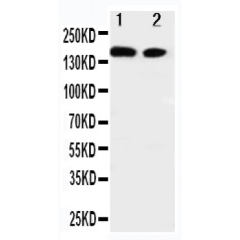
Anti-ErbB 4 antibody, ASA-B0664, Western blotting
Lane 1: A549 Cell Lysate
Lane 2: HELA Cell Lysate
Lane 1: A549 Cell Lysate
Lane 2: HELA Cell Lysate
Anti-ErbB 4 antibody, ASA-B0664, Western blotting
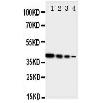
Recombinant Protein Detection Source: E.coli derived -recombinant human ERRB4, 40.6KD(162aa tag+M1-P200)
Lane 1: Recombinant Human ERRB4 Protein 10ng
Lane 2: Recombinant Human ERRB4 Protein 5ng
Lane 3: Recombinant Human ERRB4 Protein 2.5ng
Lane 4: Recombinant Human ERRB4 Protein 1.25ng
Recombinant Protein Detection Source: E.coli derived -recombinant human ERRB4, 40.6KD(162aa tag+M1-P200)
Lane 1: Recombinant Human ERRB4 Protein 10ng
Lane 2: Recombinant Human ERRB4 Protein 5ng
Lane 3: Recombinant Human ERRB4 Protein 2.5ng
Lane 4: Recombinant Human ERRB4 Protein 1.25ng
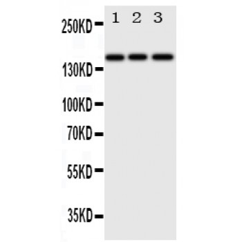
Anti-ErbB 4 antibody, ASA-B0664, Western blotting
Lane 1: HELA Cell Lysate
Lane 2: U87 Cell Lysate
Lane 3: NEURO Cell Lysate
Lane 1: HELA Cell Lysate
Lane 2: U87 Cell Lysate
Lane 3: NEURO Cell Lysate