More info
Overview
Long Name | Antibody Type | Antibody Isotype | Host | Species Reactivity | Validated Applications | Purification |
| epidermal growth factor receptor | Polyclonal | IgG | Rabbit | Human, Rat | WB | Immunogen affinity purified. |
Immunogen | ||||||
| E.coli-derived human EGFR recombinant protein (Position: L25-K346). Human EGFR shares 89% amino acid (aa) sequence identity with mouse EGFR. | ||||||
Properties
Form | Lyophilized |
Size | 100 µg/vial |
Contents | Antibody is lyophilized with 5 mg BSA, 0.9 mg NaCl, 0.2 mg Na2HPO4, 0.05 mg NaN3. *carrier free antibody available upon request. |
Concentration | Reconstitute with 0.2 mL sterile dH2O (500 µg/ml final concentration). |
Storage | At -20 °C for 12 months, as supplied. Store reconstituted antibody at 2-8 °C for one month. For long-term storage, aliquot and store at -20 °C. Avoid repeated freezing and thawing. |
Additional Information Regarding the Antigen
Gene | EGFR |
Protein | Epidermal growth factor receptor |
Uniprot ID | P00533 |
Function | Receptor tyrosine kinase binding ligands of the EGF family and activating several signaling cascades to convert extracellular cues into appropriate cellular responses. Known ligands include EGF, TGFA/TGF-alpha, amphiregulin, epigen/EPGN, BTC/betacellulin, epiregulin/EREG and HBEGF/heparin-binding EGF. Ligand binding triggers receptor homo- and/or heterodimerization and autophosphorylation on key cytoplasmic residues. The phosphorylated receptor recruits adapter proteins like GRB2 which in turn activates complex downstream signaling cascades. Activates at least 4 major downstream signaling cascades including the RAS- RAF-MEK-ERK, PI3 kinase-AKT, PLCgamma-PKC and STATs modules. May also activate the NF-kappa-B signaling cascade. Also directly phosphorylates other proteins like RGS16, activating its GTPase activity and probably coupling the EGF receptor signaling to the G protein-coupled receptor signaling. Also phosphorylates MUC1 and increases its interaction with SRC and CTNNB1/beta-catenin. |
Tissue Specificity | Ubiquitously expressed. Isoform 2 is also expressed in ovarian cancers. |
Sub-cellular localization | Cell membrane; Single-pass type I membrane protein. Endoplasmic reticulum membrane; Single-pass type I membrane protein. Golgi apparatus membrane; Single-pass type I membrane protein. Nucleus membrane; Single-pass type I membrane protein. Endosome. Endosome membrane. Nucleus. Note: In response to EGF, translocated from the cell membrane to the nucleus via Golgi and ER. Endocytosed upon activation by ligand. Colocalized with GPER1 in the nucleus of estrogen agonist-induced cancer-associated fibroblasts (CAF). |
Sequence Similarities | Belongs to the protein kinase superfamily. Tyr protein kinase family. EGF receptor subfamily. |
Aliases | Avian erythroblastic leukemia viral (v erb b) oncogene homolog antibody|Avian erythroblastic leukemia viral (verbb) oncogene homolog antibody|Cell growth inhibiting protein 40 antibody|Cell proliferation inducing protein 61 antibody|EGF R antibody|EGFR antibody|EGFR_HUMAN antibody|Epidermal growth factor receptor (avian erythroblastic leukemia viral (v erb b) oncogene homolog) antibody|Epidermal growth factor receptor (erythroblastic leukemia viral (v erb b) oncogene homolog avian) antibody|Epidermal growth factor receptor antibody|erbb 1 antibody|Erbb antibody|Erbb1 antibody|ERBB1 antibody|Errp antibody|HER1 antibody|mENA antibody|Oncogene ERBB antibody|PIG61 antibody|Proto-oncogene c-ErbB-1 antibody|Receptor tyrosine protein kinase ErbB 1 antibody|Receptor tyrosine protein kinase ErbB1 antibody|Receptor tyrosine-protein kinase ErbB-1 antibody|Urogastrone antibody|wa2 antibody|Wa5 antibody |
Application Details
| Application | Concentration* | Species | Validated Using** |
| Western blot | 0.1-0.5μg/ml | Human, Rat | AssaySolutio's ECL kit |
AssaySolution recommends Rabbit Chemiluminescent WB Detection Kit (AKIT001B) for Western blot. *Blocking peptide can be purchased at $65. Contact us for more information

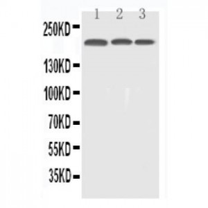
Anti-EGFR antibody, ASA-B0636--1.jpg
All lanes: Anti-EGFR(ASA-B0636) at 0.5ug/ml
Lane 1: HELA Whole Cell Lysate at 40ug
Lane 2: A549 Whole Cell Lysate at 40ug
Lane 3: A431 Whole Cell Lysate at 40ug
Predicted bind size: 134KD
Observed bind size: 200KD
All lanes: Anti-EGFR(ASA-B0636) at 0.5ug/ml
Lane 1: HELA Whole Cell Lysate at 40ug
Lane 2: A549 Whole Cell Lysate at 40ug
Lane 3: A431 Whole Cell Lysate at 40ug
Predicted bind size: 134KD
Observed bind size: 200KD


