

More info
Overview
Long Name |
Antibody Type |
Antibody Isotype |
Host |
Species Reactivity |
Validated Applications |
Purification |
| insulin receptor | Polyclonal | IgG | Rabbit | Human, Mouse, Rat | WB | Immunogen affinity purified. |
Immunogen |
||||||
| A synthetic peptide corresponding to a sequence at the C-terminus of human Insulin Receptor(1353-1372aa RSYEEHIPYTHMNGGKKNGR), different from the related rat and mouse sequences by two amino acids. | ||||||
Additional Information Regarding the Antigen
| Gene ID | 3643 |
| 3643 | P06213 |
Storage |
-20℃. Avoid freeze / thaw cycles. |
Buffer |
PBS with 0.02% sodium azide, 50% glycerol, pH 7.3. |
Gene |
INSR |
Protein |
Insulin receptor(IR) |
Uniprot ID |
P06213 |
Function |
Receptor tyrosine kinase which mediates the pleiotropic actions of insulin. Binding of insulin leads to phosphorylation of several intracellular substrates, including, insulin receptor substrates (IRS1, 2, 3, 4), SHC, GAB1, CBL and other signaling intermediates. Each of these phosphorylated proteins serve as docking proteins for other signaling proteins that contain Src- homology-2 domains (SH2 domain) that specifically recognize different phosphotyrosines residues, including the p85 regulatory subunit of PI3K and SHP2. Phosphorylation of IRSs proteins lead to the activation of two main signaling pathways: the PI3K-AKT/PKB pathway, which is responsible for most of the metabolic actions of insulin, and the Ras-MAPK pathway, which regulates expression of some genes and cooperates with the PI3K pathway to control cell growth and differentiation. Binding of the SH2 domains of PI3K to phosphotyrosines on IRS1 leads to the activation of PI3K and the generation of phosphatidylinositol-(3, 4, 5)-triphosphate (PIP3), a lipid second messenger, which activates several PIP3-dependent serine/threonine kinases, such as PDPK1 and subsequently AKT/PKB. The net effect of this pathway is to produce a translocation of the glucose transporter SLC2A4/GLUT4 from cytoplasmic vesicles to the cell membrane to facilitate glucose transport. Moreover, upon insulin stimulation, activated AKT/PKB is responsible for: anti- apoptotic effect of insulin by inducing phosphorylation of BAD; regulates the expression of gluconeogenic and lipogenic enzymes by controlling the activity of the winged helix or forkhead (FOX) class of transcription factors. Another pathway regulated by PI3K- AKT/PKB activation is mTORC1 signaling pathway which regulates cell growth and metabolism and integrates signals from insulin. AKT mediates insulin-stimulated protein synthesis by phosphorylating TSC2 thereby activating mTORC1 pathway. The Ras/RAF/MAP2K/MAPK pathway is mainly involved in mediating cell growth, survival and cellular differentiation of insulin. Phosphorylated IRS1 recruits GRB2/SOS complex, which triggers the activation of the Ras/RAF/MAP2K/MAPK pathway. In addition to binding insulin, the insulin receptor can bind insulin-like growth factors (IGFI and IGFII). Isoform Short has a higher affinity for IGFII binding. When present in a hybrid receptor with IGF1R, binds IGF1. PubMed:12138094 shows that hybrid receptors composed of IGF1R and INSR isoform Long are activated with a high affinity by IGF1, with low affinity by IGF2 and not significantly activated by insulin, and that hybrid receptors composed of IGF1R and INSR isoform Short are activated by IGF1, IGF2 and insulin. In contrast, PubMed:16831875 shows that hybrid receptors composed of IGF1R and INSR isoform Long and hybrid receptors composed of IGF1R and INSR isoform Short have similar binding characteristics, both bind IGF1 and have a low affinity for insulin. |
Tissue Specificity |
Isoform Long and isoform Short are predominantly expressed in tissue targets of insulin metabolic effects: liver, adipose tissue and skeletal muscle but are also expressed in the peripheral nerve, kidney, pulmonary alveoli, pancreatic acini, placenta vascular endothelium, fibroblasts, monocytes, granulocytes, erythrocytes and skin. Isoform Short is preferentially expressed in fetal cells such as fetal fibroblasts, muscle, liver and kidney. Found as a hybrid receptor with IGF1R in muscle, heart, kidney, adipose tissue, skeletal muscle, hepatoma, fibroblasts, spleen and placenta (at protein level). Overexpressed in several tumors, including breast, colon, lung, ovary, and thyroid carcinomas. |
Sub-cellular localization |
Cell membrane; Single-pass type I membrane protein. |
Sequence Similarities |
Belongs to the protein kinase superfamily. Tyr protein kinase family. Insulin receptor subfamily. |
Aliases |
CD 220 antibody|CD220 antibody|CD220 antigen antibody|HHF 5 antibody|HHF5 antibody|human insulin receptor antibody|INSR antibody|INSR_HUMAN antibody|Insulin receptor subunit beta antibody|IR 1 antibody|IR antibody|IR-1 antibody|IR1 antibody |
Application Details
| Application | Concentration* | Species | Validated Using** |
| Western blot | 0.1-0.5μg/ml | Human, Rat Mouse | AssaySolutio's ECL kit |
AssaySolution recommends Rabbit Chemiluminescent WB Detection Kit (AKIT001B) for Western blot. *Blocking peptide can be purchased at $65. Contact us for more information
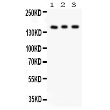
Anti- Insulin Receptor antibody, ASA-B1039, Western blotting
All lanes: Anti Insulin Receptor(ASA-B1039) at 0.5ug/ml
Lane 1: Rat Kidney Tissue Lysate at 50ug
Lane 2: PANC Whole Cell Lysate at 40ug
Lane 3: HELA Whole Cell Lysate at 40ug
Predicted bind size: 155KD
Observed bind size: 155KD
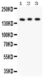
All lanes: Anti Insulin Receptor(ASA-B1039) at 0.5ug/ml
Lane 1: Rat Kidney Tissue Lysate at 50ug
Lane 2: PANC Whole Cell Lysate at 40ug
Lane 3: HELA Whole Cell Lysate at 40ug
Predicted bind size: 155KD
Observed bind size: 155KD
|
Gene ID |
|
|
Swiss prot |
|
|
Storage |
Store at -20℃. Avoid freeze / thaw cycles. Buffer: PBS with 0.02% sodium azide, 50% glycerol, pH7.3. |