

More info
Overview
Long Name | Antibody Type | Antibody Isotype | Host | Species Reactivity | Validated Applications | Purification |
| SMAD family member 3 | Polyclonal | IgG | Rabbit | Human | WB | Immunogen affinity purified. |
Immunogen | ||||||
| A synthetic peptide corresponding to a sequence in the middle region of human Smad3 (193-222aa DHQMNHSMDAGSPNLSPNPMSPAHNNLDLQ), identical to the related mouse and rat sequences. | ||||||
Properties
Form | Lyophilized |
Size | 100 µg/vial |
Contents | Antibody is lyophilized with 5 mg BSA, 0.9 mg NaCl, 0.2 mg Na2HPO4, 0.05 mg NaN3. *carrier free antibody available upon request. |
Concentration | Reconstitute with 0.2 mL sterile dH2O (500 µg/ml final concentration). |
Storage | At -20 °C for 12 months, as supplied. Store reconstituted antibody at 2-8 °C for one month. For long-term storage, aliquot and store at -20 °C. Avoid repeated freezing and thawing. |
Additional Information Regarding the Antigen
Gene | SMAD3 |
Protein | Mothers against decapentaplegic homolog 3 |
Uniprot ID | P84022 |
Function | Receptor-regulated SMAD (R-SMAD) that is an intracellular signal transducer and transcriptional modulator activated by TGF-beta (transforming growth factor) and activin type 1 receptor kinases. Binds the TRE element in the promoter region of many genes that are regulated by TGF-beta and, on formation of the SMAD3/SMAD4 complex, activates transcription. Also can form a SMAD3/SMAD4/JUN/FOS complex at the AP-1/SMAD site to regulate TGF-beta-mediated transcription. Has an inhibitory effect on wound healing probably by modulating both growth and migration of primary keratinocytes and by altering the TGF- mediated chemotaxis of monocytes. This effect on wound healing appears to be hormone-sensitive. Regulator of chondrogenesis and osteogenesis and inhibits early healing of bone fractures. Positively regulates PDPK1 kinase activity by stimulating its dissociation from the 14-3-3 protein YWHAQ which acts as a negative regulator. |
Tissue Specificity | |
Sub-cellular localization | Cytoplasm. Nucleus. Note: Cytoplasmic and nuclear in the absence of TGF-beta. On TGF-beta stimulation, migrates to the nucleus when complexed with SMAD4. Through the action of the phosphatase PPM1A, released from the SMAD2/SMAD4 complex, and exported out of the nucleus by interaction with RANBP1. Co-localizes with LEMD3 at the nucleus inner membrane. MAPK-mediated phosphorylation appears to have no effect on nuclear import. PDPK1 prevents its nuclear translocation in response to TGF-beta. |
Sequence Similarities | Belongs to the dwarfin/SMAD family. |
Aliases | DKFZP586N0721 antibody|DKFZp686J10186 antibody|hMAD 3 antibody| hMAD-3 antibody|hSMAD3 antibody|HSPC193 antibody|HST17436 antibody| JV15 2 antibody|JV15-2 antibody|JV152 antibody|JV152 antibody|LDS1C antibody|LDS3 antibody|MAD (mothers against decapentaplegic Drosophila) homolog 3 antibody|MAD (mothers against decapentaplegic Drosophila) homolog 3 antibody|MAD homolog 3 antibody|Mad homolog JV15 2 antibody| Mad protein homolog antibody|MAD, mothers against decapentaplegic homolog 3 antibody|MAD3 antibody|MAD3 antibody|MADH 3 antibody|MADH3 antibody|MADH3 antibody|MGC60396 antibody|Mothers against decapentaplegic homolog 3 antibody|Mothers against DPP homolog 3 antibody|Mothers against DPP homolog 3 antibody|SMA and MAD related protein 3 antibody|SMAD 3 antibody|SMAD antibody|SMAD family member 3 antibody|SMAD, mothers against DPP homolog 3 antibody|SMAD3 antibody| SMAD3_HUMAN antibody |
Application Details
| Application | Concentration* | Species | Validated Using** |
| Western blot | 0.1-0.5μg/ml | Human | AssaySolutio's ECL kit |
AssaySolution recommends Rabbit Chemiluminescent WB Detection Kit (AKIT001B) for Western blot. *Blocking peptide can be purchased at $65. Contact us for more information

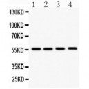
Anti- SMAD3 antibody, ASA-B1729, Western blotting
All lanes: Anti SMAD3 (ASA-B1729) at 0.5ug/ml
Lane 1: U87 Whole Cell Lysate at 40ug
Lane 2: Human Placenta Tissue Lysate at 50ug
Lane 3: PANC Whole Cell Lysate at 40ug
Lane 4: HELA Whole Cell Lysate at 40ug
Predicted bind size: 48KD
Observed bind size: 56KD
All lanes: Anti SMAD3 (ASA-B1729) at 0.5ug/ml
Lane 1: U87 Whole Cell Lysate at 40ug
Lane 2: Human Placenta Tissue Lysate at 50ug
Lane 3: PANC Whole Cell Lysate at 40ug
Lane 4: HELA Whole Cell Lysate at 40ug
Predicted bind size: 48KD
Observed bind size: 56KD
