

More info
Overview
Long Name | Antibody Type | Antibody Isotype | Host | Species Reactivity | Validated Applications | Purification |
| aurora kinase A | Polyclonal | IgG | Rabbit | Mouse, Rat | WB | Immunogen affinity purified. |
Immunogen | ||||||
| A synthetic peptide corresponding to a sequence in the middle region of mouse Aurora A(109-125aa QKTEDTKKRQWTLEDFD), different from the related rat sequence by one amino acid. | ||||||
Properties
Form | Lyophilized |
Size | 100 µg/vial |
Contents | Antibody is lyophilized with 5 mg BSA, 0.9 mg NaCl, 0.2 mg Na2HPO4, 0.05 mg Thimerosal and 0.05 mg NaN3. *carrier free antibody available upon request. |
Concentration | Reconstitute with 0.2 mL sterile dH2O (500 µg/ml final concentration). |
Storage | At -20 °C for 12 months, as supplied. Store reconstituted antibody at 2-8 °C for one month. For long-term storage, aliquot and store at -20 °C. Avoid repeated freezing and thawing. |
Additional Information Regarding the Antigen
Gene | AURKA |
Protein | Aurora kinase A |
Uniprot ID | P97477 |
Function | Mitotic serine/threonine kinases that contributes to the regulation of cell cycle progression. Associates with the centrosome and the spindle microtubules during mitosis and plays a critical role in various mitotic events including the establishment of mitotic spindle, centrosome duplication, centrosome separation as well as maturation, chromosomal alignment, spindle assembly checkpoint, and cytokinesis. Required for initial activation of CDK1 at centrosomes. Phosphorylates numerous target proteins, including ARHGEF2, BORA, BRCA1, CDC25B, DLGP5, HDAC6, KIF2A, LATS2, NDEL1, PARD3, PPP1R2, PLK1, RASSF1, TACC3, p53/TP53 and TPX2. Regulates KIF2A tubulin depolymerase activity. Required for normal axon formation. Plays a role in microtubule remodeling during neurite extension. Important for microtubule formation and/or stabilization. Also acts as a key regulatory component of the p53/TP53 pathway, and particularly the checkpoint-response pathways critical for oncogenic transformation of cells, by phosphorylating and stabilizing p53/TP53. Phosphorylates its own inhibitors, the protein phosphatase type 1 (PP1) isoforms, to inhibit their activity. Necessary for proper cilia disassembly prior to mitosis. |
Tissue Specificity | Detected in embryonic neurons in dorsal root ganglia and brain cortex (at protein level). Highly expressed in testis, in about one third of the seminiferous tubules. Expression is restricted to specific spermatocytes nearing completion of prophase, with levels falling off on transition to elongated spermatids. Highly expressed in the ovary, expression in the oocyte starts around the transition to large growing follicle. Abundant expression is seen in the proliferating granulosa and thecal cells of the growing follicle, and in the young corpus luteum. Very weakly expressed in spleen and intestine. |
Sub-cellular localization | Cytoplasm, cytoskeleton, microtubule organizing center, centrosome. Cytoplasm, cytoskeleton, spindle pole. Note: Localizes on centrosomes in interphase cells and at each spindle pole in mitosis. Associates with both the pericentriolar material (PCM) and centrioles. Colocalized with SIRT2 at centrosome (By similarity). Detected at the neurite hillock in developing neurons. |
Sequence Similarities | Belongs to the protein kinase superfamily. Ser/Thr protein kinase family. Aurora subfamily. |
Aliases | AIK antibody|ARK-1 antibody|ARK1 antibody|AURA antibody|AURKA antibody|Aurora 2 antibody|Aurora A antibody|Aurora Family Kinase 1 antibody|aurora kinase A antibody|Aurora Related Kinase 1 antibody|Aurora-related kinase 1 antibody|Aurora/IPL1 Like Kinase antibody|Aurora/IPL1 Related Kinase 1 antibody|Aurora/IPL1-related kinase 1 antibody|AURORA2 antibody|AYK1 antibody|BRAK antibody|Breast Tumor Amplified Kinase antibody|Breast tumor-amplified kinase antibody|BTAK antibody|hARK1 antibody|IAK antibody|IPL1 Aurora Related Kinase 1 antibody|IPL1 Related Kinase antibody|MGC34538 antibody|OTTHUMP00000031340 antibody|OTTHUMP00000031341 antibody|OTTHUMP00000031342 antibody|OTTHUMP00000031343 antibody|OTTHUMP00000031344 antibody|OTTHUMP00000031345 antibody|OTTHUMP00000166071 antibody|OTTHUMP00000166072 antibody|PPP1R47 antibody|Protein phosphatase 1, regulatory subunit 47 antibody|Serine/threonine kinase 15 antibody|Serine/threonine kinase 6 antibody|Serine/threonine kinase antibody|Serine/threonine-protein kinase 15 antibody|Serine/threonine-protein kinase 6 antibody|Serine/threonine-protein kinase aurora-A antibody|STK15 antibody|STK6 antibody|STK6_HUMAN antibody|STK7 antibody |
Application Details
| Application | Concentration* | Species | Validated Using** |
| Western blot | 0.1-0.5μg/ml | Mouse Rat | AssaySolutio's ECL kit |
AssaySolution recommends Rabbit Chemiluminescent WB Detection Kit (AKIT001B) for Western blot. *Blocking peptide can be purchased at $65. Contact us for more information
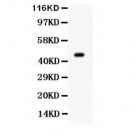
Anti- AURKA antibody, ASA-B0179, Western blotting
Anti AURKA (ASA-B0179) at 0.5ug/ml
Mouse Ovary Tissue Lysate at 50ug
Predicted band size: 46KD
Observed band size: 46KD
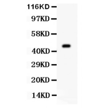
Anti AURKA (ASA-B0179) at 0.5ug/ml
Mouse Ovary Tissue Lysate at 50ug
Predicted band size: 46KD
Observed band size: 46KD