More info
Overview
Long Name | Antibody Type | Antibody Isotype | Host | Species Reactivity | Validated Applications | Purification |
| ubiquitin D | Polyclonal | IgG | Rabbit | Human | IHC-P, WB | Immunogen affinity purified. |
Immunogen | ||||||
| A synthetic peptide corresponding to a sequence at the N-terminus of human Diubiquitin(27-40aa YDSVKKIKEHVRSK), different from the related mouse and rat sequences by five amino acids. | ||||||
Properties
Form | Lyophilized |
Size | 100 µg/vial |
Contents | Antibody is lyophilized with 5 mg BSA, 0.9 mg NaCl, 0.2 mg Na2HPO4, 0.05 mg Thimerosal and 0.05 mg NaN3. *carrier free antibody available upon request. |
Concentration | Reconstitute with 0.2 mL sterile dH2O (500 µg/ml final concentration). |
Storage | At -20 °C for 12 months, as supplied. Store reconstituted antibody at 2-8 °C for one month. For long-term storage, aliquot and store at -20 °C. Avoid repeated freezing and thawing. |
Additional Information Regarding the Antigen
Gene | UBD |
Protein | Ubiquitin D |
Uniprot ID | O15205 |
Function | Ubiquitin-like protein modifier which can be covalently attached to target protein and subsequently leads to their degradation by the 26S proteasome, in a NUB1L-dependent manner. Probably functions as a survival factor. Conjugation ability activated by UBA6. Promotes the expression of the proteasome subunit beta type-9 (PSMB9/LMP2). Regulates TNF-alpha-induced and LPS-mediated activation of the central mediator of innate immunity NF-kappa-B by promoting TNF-alpha-mediated proteasomal degradation of ubiquitinated-I-kappa-B-alpha. Required for TNF-alpha-induced p65 nuclear translocation in renal tubular epithelial cells (RTECs). May be involved in dendritic cell (DC) maturation, the process by which immature dendritic cells differentiate into fully competent antigen-presenting cells that initiate T-cell responses. Mediates mitotic non-disjunction and chromosome instability, in long-term in vitro culture and cancers, by abbreviating mitotic phase and impairing the kinetochore localization of MAD2L1 during the prometaphase stage of the cell cycle. May be involved in the formation of aggresomes when proteasome is saturated or impaired. Mediates apoptosis in a caspase-dependent manner, especially in renal epithelium and tubular cells during renal diseases such as polycystic kidney disease and Human immunodeficiency virus (HIV)- associated nephropathy (HIVAN). |
Tissue Specificity | Constitutively expressed in mature dendritic cells and B-cells. Mostly expressed in the reticuloendothelial system (e.g. thymus, spleen), the gastrointestinal system, kidney, lung and prostate gland. |
Sub-cellular localization | Nucleus. |
Sequence Similarities | Contains 2 ubiquitin-like domains. |
Aliases | Diubiquitin antibody|FAT10 antibody|UBD 3 antibody|Ubd antibody|UBD_HUMAN antibody|Ubiquitin D antibody|Ubiquitin like protein FAT10 antibody|Ubiquitin-like protein FAT10 antibody |
Application Details
| Application | Concentration* | Species | Validated Using** |
| Western blot | 0.1-0.5μg/ml | Human | AssaySolutio's ECL kit |
| Immunohistochemistry(Paraffin-embedded Section) | 0.5-1μg/ml | Human | AssaySolutio's IHC/ICC Detection kit |
AssaySolution recommends Rabbit Chemiluminescent WB Detection Kit (AKIT001B) for Western blot, and Rabbit Peroxidase IHC/ICC Detection Kit (AKIT002B) for IHC(P). *Blocking peptide can be purchased at $65. Contact us for more information

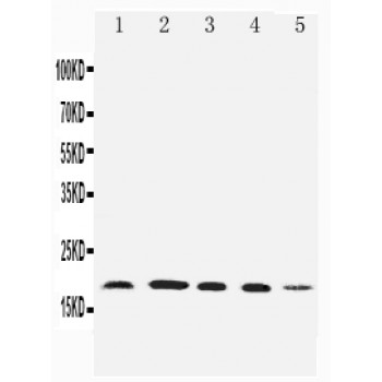
Anti-Diubiquitin antibody, ASA-B0593, Western blotting
Lane 1: HELA Cell Lysate
Lane 2: SKOV Cell Lysate
Lane 3: MCF-7 Cell Lysate
Lane 4: A549 Cell Lysate
Lane 5: SMMC Cell Lysate
Lane 1: HELA Cell Lysate
Lane 2: SKOV Cell Lysate
Lane 3: MCF-7 Cell Lysate
Lane 4: A549 Cell Lysate
Lane 5: SMMC Cell Lysate

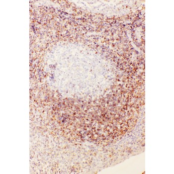
Anti-Diubiquitin antibody, ASA-B0593, IHC(P)
IHC(P): Human Tonsil Tissue
IHC(P): Human Tonsil Tissue

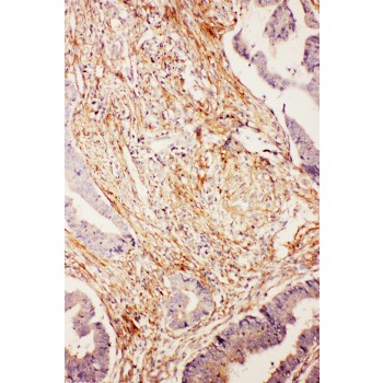
Anti-Diubiquitin antibody, ASA-B0593, IHC(P)
IHC(P): Human Intestinal Cancer Tissue
IHC(P): Human Intestinal Cancer Tissue


