

More info
Overview
Long Name | Antibody Type | Antibody Isotype | Host | Species Reactivity | Validated Applications | Purification |
| leupaxin | Polyclonal | IgG | Rabbit | Human, Mouse, Rat | IHC-P, WB | Immunogen affinity purified. |
Immunogen | ||||||
| A synthetic peptide corresponding to a sequence at the N-terminus of human Leupaxin(115-129aa KKHLPDKQDHKASLD), different from the related rat sequence by two amino acids, and from the related mouse sequence by three amino acids. | ||||||
Properties
Form | Lyophilized |
Size | 100 µg/vial |
Contents | Antibody is lyophilized with 5 mg BSA, 0.9 mg NaCl, 0.2 mg Na2HPO4, 0.05 mg Thimerosal and 0.05 mg NaN3. *carrier free antibody available upon request. |
Concentration | Reconstitute with 0.2 mL sterile dH2O (500 µg/ml final concentration). |
Storage | At -20 °C for 12 months, as supplied. Store reconstituted antibody at 2-8 °C for one month. For long-term storage, aliquot and store at -20 °C. Avoid repeated freezing and thawing. |
Additional Information Regarding the Antigen
Gene | LPXN |
Protein | Leupaxin |
Uniprot ID | O60711 |
Function | Transcriptional coactivator for androgen receptor (AR) and serum response factor (SRF). Contributes to the regulation of cell adhesion, spreading and cell migration and acts as a negative regulator in integrin-mediated cell adhesion events. Suppresses the integrin-induced tyrosine phosphorylation of paxillin (PXN). May play a critical role as an adapter protein in the formation of the adhesion zone in osteoclasts. Negatively regulates B-cell antigen receptor (BCR) signaling. |
Tissue Specificity | Macrophages, monocytes and osteoclasts (at protein level). Strongly expressed in cells and tissues of hematopoietic origin. Highest expression in lymphoid tissues such as spleen, lymph node, thymus and appendix and in the vascular smooth muscle. Lower levels in bone marrow and fetal liver. Also expressed in peripheral blood lymphocytes and a number of hematopoietic cell lines. Very low levels found in epithelial cell lines. Expressed in prostate cancer (PCa) cells and its expression intensity is directly linked to PCa progression. |
Sub-cellular localization | Cytoplasm. Cell junction, focal adhesion. Nucleus. Cytoplasm, perinuclear region . Cell projection, podosome. Cell membrane. Note: Shuttles between the cytoplasm and nucleus. Recruited to the cell membrane following B- cell antigen receptor (BCR) cross-linking in B-cells. Enhanced focal adhesion kinase activity (PTK2/FAK) attenuates its nuclear accumulation and limits its ability to enhance serum response factor (SRF)-dependent gene transcription. Targeting to focal adhesions is essential for its tyrosine phosphorylation in response to bombesin. |
Sequence Similarities | Belongs to the paxillin family. |
Aliases | LDLP antibody|LDPL antibody|LPXN antibody |
Application Details
| Application | Concentration* | Species | Validated Using** |
| Western blot | 0.1-0.5μg/ml | Human, Rat | AssaySolutio's ECL kit |
| Immunohistochemistry(Paraffin-embedded Section) | 0.5-1μg/ml | Human, Mouse, Rat | AssaySolutio's IHC/ICC Detection kit |
AssaySolution recommends Rabbit Chemiluminescent WB Detection Kit (AKIT001B) for Western blot, and Rabbit Peroxidase IHC/ICC Detection Kit (AKIT002B) for IHC(P). *Blocking peptide can be purchased at $65. Contact us for more information
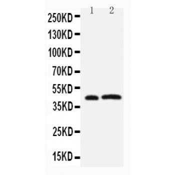
Anti-Leupaxin antibody, ASA-B1166, Western blotting
Lane 1: Rat Thymus Tissue Lysate
Lane 2: JURKAT Cell Lysate
Lane 1: Rat Thymus Tissue Lysate
Lane 2: JURKAT Cell Lysate
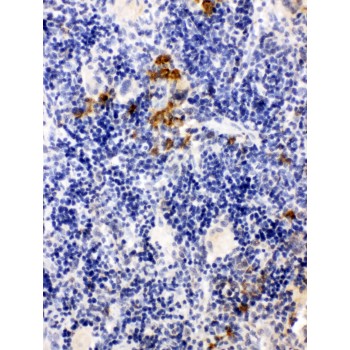
Anti-Leupaxin antibody, ASA-B1166, IHC(P)
IHC(P): Rat Spleen Tissue
IHC(P): Rat Spleen Tissue
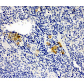
Anti-Leupaxin antibody, ASA-B1166, IHC(P)
IHC(P): Mouse Spleen Tissue
IHC(P): Mouse Spleen Tissue