More info
Overview
Long Name | Antibody Type | Antibody Isotype | Host | Species Reactivity | Validated Applications | Purification |
| inositol polyphosphate phosphatase-like 1 | Polyclonal | IgG | Rabbit | Human, Mouse, Rat | IHC-P, WB | Immunogen affinity purified. |
Immunogen | ||||||
| A synthetic peptide corresponding to a sequence in the middle region of human INPPL1(349-363aa QRDSQEDWTTFTHDR), identical to the related rat and mouse sequences. | ||||||
Properties
Form | Lyophilized |
Size | 100 µg/vial |
Contents | Antibody is lyophilized with 5 mg BSA, 0.9 mg NaCl, 0.2 mg Na2HPO4, 0.05 mg Thimerosal and 0.05 mg NaN3. *carrier free antibody available upon request. |
Concentration | Reconstitute with 0.2 mL sterile dH2O (500 µg/ml final concentration). |
Storage | At -20 °C for 12 months, as supplied. Store reconstituted antibody at 2-8 °C for one month. For long-term storage, aliquot and store at -20 °C. Avoid repeated freezing and thawing. |
Additional Information Regarding the Antigen
Gene | INPPL1 |
Protein | Phosphatidylinositol 3,4,5-trisphosphate 5-phosphatase 2 |
Uniprot ID | O15357 |
Function | Phosphatidylinositol (PtdIns) phosphatase that specifically hydrolyzes the 5-phosphate of phosphatidylinositol- 3,4,5-trisphosphate (PtdIns(3,4,5)P3) to produce PtdIns(3,4)P2, thereby negatively regulating the PI3K (phosphoinositide 3-kinase) pathways. Plays a central role in regulation of PI3K-dependent insulin signaling, although the precise molecular mechanisms and signaling pathways remain unclear. While overexpression reduces both insulin-stimulated MAP kinase and Akt activation, its absence does not affect insulin signaling or GLUT4 trafficking. Confers resistance to dietary obesity. May act by regulating AKT2, but not AKT1, phosphorylation at the plasma membrane. Part of a signaling pathway that regulates actin cytoskeleton remodeling. Required for the maintenance and dynamic remodeling of actin structures as well as in endocytosis, having a major impact on ligand-induced EGFR internalization and degradation. Participates in regulation of cortical and submembraneous actin by hydrolyzing PtdIns(3,4,5)P3 thereby regulating membrane ruffling. Regulates cell adhesion and cell spreading. Required for HGF-mediated lamellipodium formation, cell scattering and spreading. Acts as a negative regulator of EPHA2 receptor endocytosis by inhibiting via PI3K-dependent Rac1 activation. Acts as a regulator of neuritogenesis by regulating PtdIns(3,4,5)P3 level and is required to form an initial protrusive pattern, and later, maintain proper neurite outgrowth. Acts as a negative regulator of the FC-gamma-RIIA receptor (FCGR2A). Mediates signaling from the FC-gamma-RIIB receptor (FCGR2B), playing a central role in terminating signal transduction from activating immune/hematopoietic cell receptor systems. Involved in EGF signaling pathway. Upon stimulation by EGF, it is recruited by EGFR and dephosphorylates PtdIns(3,4,5)P3. Plays a negative role in regulating the PI3K-PKB pathway, possibly by inhibiting PKB activity. Down-regulates Fc-gamma-R-mediated phagocytosis in macrophages independently of INPP5D/SHIP1. In macrophages, down-regulates NF-kappa-B-dependent gene transcription by regulating macrophage colony-stimulating factor (M-CSF)-induced signaling. May also hydrolyze PtdIns(1,3,4,5)P4, and could thus affect the levels of the higher inositol polyphosphates like InsP6. Involved in endochondral ossification. |
Tissue Specificity | Widely expressed, most prominently in skeletal muscle, heart and brain. Present in platelets. Expressed in transformed myeloid cells and in primary macrophages, but not in peripheral blood monocytes. |
Sub-cellular localization | Cytoplasm, cytosol. Cytoplasm, cytoskeleton. Membrane; Peripheral membrane protein. Cell projection, filopodium. Cell projection, lamellipodium. Note: Translocates to membrane ruffles when activated, translocation is probably due to different mechanisms depending on the stimulus and cell type. Partly translocated via its SH2 domain which mediates interaction with tyrosine phosphorylated receptors such as the FC-gamma-RIIB receptor (FCGR2B). Tyrosine phosphorylation may also participate in membrane localization. Insulin specifically stimulates its redistribution from the cytosol to the plasma membrane. Recruited to the membrane following M-CSF stimulation. In activated spreading platelets, localizes with actin at filopodia, lamellipodia and the central actin ring. |
Sequence Similarities | Belongs to the inositol 1,4,5-trisphosphate 5- phosphatase family. |
Aliases | 4 antibody|5-trisphosphate 5-phosphatase 2 antibody|51C protein antibody|EC 3.1.3.n1 antibody|inositol polyphosphate phosphatase like 1 antibody|Inositol polyphosphate phosphatase like protein 1 antibody|Inositol polyphosphate phosphatase-like protein 1 antibody|INPPL-1 antibody|INPPL1 antibody|Phosphatidylinositol 3 antibody|Phosphatidylinositol 3,4,5 trisphosphate 5 phosphatase 2 antibody|Protein 51C antibody|SH2 domain containing inositol 5' phosphatase 2 antibody|SH2 domain-containing inositol 5''-phosphatase 2 antibody|SH2 domain-containing inositol phosphatase 2 antibody|SHIP-2 antibody|SHIP2 antibody|SHIP2_HUMAN antibody |
Application Details
| Application | Concentration* | Species | Validated Using** |
| Western blot | 0.1-0.5μg/ml | Human, Rat Mouse | AssaySolutio's ECL kit |
| Immunohistochemistry(Paraffin-embedded Section) | 0.5-1μg/ml | Human Mouse, Rat | AssaySolutio's IHC/ICC Detection kit |
AssaySolution recommends Rabbit Chemiluminescent WB Detection Kit (AKIT001B) for Western blot, and Rabbit Peroxidase IHC/ICC Detection Kit (AKIT002B) for IHC(P). *Blocking peptide can be purchased at $65. Contact us for more information
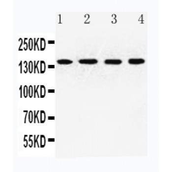
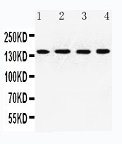
Anti-INPPL1 antibody, ASA-B1036, Western blotting
Lane 1: Rat Heart Tissue Lysate
Lane 2: U87 Cell Lysate
Lane 3: HT1080 Cell Lysate
Lane 4: MCF-7 Cell Lysate
Lane 1: Rat Heart Tissue Lysate
Lane 2: U87 Cell Lysate
Lane 3: HT1080 Cell Lysate
Lane 4: MCF-7 Cell Lysate
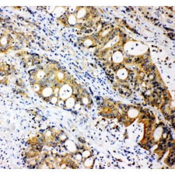
Anti-INPPL1 antibody, ASA-B1036, IHC(P)
IHC(P): Human Intestinal Cancer Tissue
IHC(P): Human Intestinal Cancer Tissue