

More info
Overview
Long Name | Antibody Type | Antibody Isotype | Host | Species Reactivity | Validated Applications | Purification |
| amyloid beta(A4) precursor protein | Polyclonal | IgG | Rabbit | Human, Mouse, Rat | IHC-P, WB | Immunogen affinity purified. |
Immunogen | ||||||
| A synthetic peptide corresponding to a sequence at the N-terminus of human Amyloid beta precursor protein(18-32aa EVPTDGNAGLLAEPQ), identical to the related mouse and rat sequences. | ||||||
Properties
Form | Lyophilized |
Size | 100 µg/vial |
Contents | Antibody is lyophilized with 5 mg BSA, 0.9 mg NaCl, 0.2 mg Na2HPO4, 0.05 mg Thimerosal and 0.05 mg NaN3. *carrier free antibody available upon request. |
Concentration | Reconstitute with 0.2 mL sterile dH2O (500 µg/ml final concentration). |
Storage | At -20 °C for 12 months, as supplied. Store reconstituted antibody at 2-8 °C for one month. For long-term storage, aliquot and store at -20 °C. Avoid repeated freezing and thawing. |
Additional Information Regarding the Antigen
Gene | APP |
Protein | Amyloid beta A4 protein |
Uniprot ID | P05067 |
Function | Functions as a cell surface receptor and performs physiological functions on the surface of neurons relevant to neurite growth, neuronal adhesion and axonogenesis. Involved in cell mobility and transcription regulation through protein-protein interactions. Can promote transcription activation through binding to APBB1-KAT5 and inhibits Notch signaling through interaction with Numb. Couples to apoptosis-inducing pathways such as those mediated by G(O) and JIP. Inhibits G(o) alpha ATPase activity (By similarity). Acts as a kinesin I membrane receptor, mediating the axonal transport of beta-secretase and presenilin 1. Involved in copper homeostasis/oxidative stress through copper ion reduction. In vitro, copper-metallated APP induces neuronal death directly or is potentiated through Cu(2+)-mediated low-density lipoprotein oxidation. Can regulate neurite outgrowth through binding to components of the extracellular matrix such as heparin and collagen I and IV. The splice isoforms that contain the BPTI domain possess protease inhibitor activity. Induces a AGER- dependent pathway that involves activation of p38 MAPK, resulting in internalization of amyloid-beta peptide and leading to mitochondrial dysfunction in cultured cortical neurons. Provides Cu(2+) ions for GPC1 which are required for release of nitric oxide (NO) and subsequent degradation of the heparan sulfate chains on GPC1. |
Tissue Specificity | Expressed in all fetal tissues examined with highest levels in brain, kidney, heart and spleen. Weak expression in liver. In adult brain, highest expression found in the frontal lobe of the cortex and in the anterior perisylvian cortex- opercular gyri. Moderate expression in the cerebellar cortex, the posterior perisylvian cortex-opercular gyri and the temporal associated cortex. Weak expression found in the striate, extra- striate and motor cortices. Expressed in cerebrospinal fluid, and plasma. Isoform APP695 is the predominant form in neuronal tissue, isoform APP751 and isoform APP770 are widely expressed in non- neuronal cells. Isoform APP751 is the most abundant form in T- lymphocytes. Appican is expressed in astrocytes. |
Sub-cellular localization | Membrane; Single-pass type I membrane protein. Membrane, clathrin-coated pit. Note: Cell surface protein that rapidly becomes internalized via clathrin-coated pits. During maturation, the immature APP (N-glycosylated in the endoplasmic reticulum) moves to the Golgi complex where complete maturation occurs (O-glycosylated and sulfated). After alpha-secretase cleavage, soluble APP is released into the extracellular space and the C-terminal is internalized to endosomes and lysosomes. Some APP accumulates in secretory transport vesicles leaving the late Golgi compartment and returns to the cell surface. Gamma-CTF(59) peptide is located to both the cytoplasm and nuclei of neurons. It can be translocated to the nucleus through association with APBB1 (Fe65). Beta-APP42 associates with FRPL1 at the cell surface and the complex is then rapidly internalized. APP sorts to the basolateral surface in epithelial cells. During neuronal differentiation, the Thr-743 phosphorylated form is located mainly in growth cones, moderately in neurites and sparingly in the cell body. Casein kinase phosphorylation can occur either at the cell surface or within a post-Golgi compartment. Associates with GPC1 in perinuclear compartments. Colocalizes with SORL1 in a vesicular pattern in cytoplasm and perinuclear regions. |
Sequence Similarities | Belongs to the APP family. |
Aliases | A4 amyloid protein antibody|A4 antibody|A4_HUMAN antibody|AAA antibody|ABETA antibody|ABPP antibody|AD 1 antibody|AD1 antibody|AICD-50 antibody|AICD-57 antibody|AICD-59 antibody|AID(50) antibody|AID(57) antibody|AID(59) antibody|Alzheimer Disease 1 antibody|Alzheimer disease amyloid protein antibody|Alzheimer's Disease Amyloid Protein antibody|Amyloid beta(A4) precursor protein antibody|Amyloid beta A4 protein antibody|Amyloid Beta A4 Protein Precursor Isoform A antibody|Amyloid beta A4 protein precursor isoform b antibody|Amyloid beta A4 protein precursor isoform c antibody|Amyloid Beta Peptide antibody|Amyloid beta protein antibody|Amyloid intracellular domain 50 antibody|Amyloid intracellular domain 57 antibody|Amyloid intracellular domain 59 antibody|Amyloid Of Aging And Alzheimer Disease antibody|APP antibody|APPI antibody|Beta amyloid peptide antibody|Beta-APP40 antibody|Beta-APP42 antibody|C31 antibody|Cerebal Vascular Amyloid Peptide antibody|Cerebral vascular amyloid peptide antibody|CTFgamma antibody|CVAP antibody|Gamma-CTF(50) antibody|Gamma-CTF(57) antibody|Gamma-CTF(59) antibody|Human mRNA For Amyloid A4 Precursor Of Alzheimer's Disease antibody|Peptidase nexin II antibody|peptidase nexin-II antibody|PN 2 antibody|PN II antibody|PN-II antibody|PN2 antibody|PNII antibody|PREA4 antibody|Protease Nexin II antibody|Protease nexin-II antibody|S-APP-alpha antibody|S-APP-beta antibody |
Application Details
| Application | Concentration* | Species | Validated Using** |
| Western blot | 0.1-0.5μg/ml | Human, Rat Mouse | AssaySolutio's ECL kit |
| Immunohistochemistry(Paraffin-embedded Section) | 0.5-1μg/ml | Human, Rat Mouse | AssaySolutio's IHC/ICC Detection kit |
AssaySolution recommends Rabbit Chemiluminescent WB Detection Kit (AKIT001B) for Western blot, and Rabbit Peroxidase IHC/ICC Detection Kit (AKIT002B) for IHC(P). *Blocking peptide can be purchased at $65. Contact us for more information
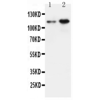
Anti-Amyloid beta precursor protein antibody, ASA-B0080, Western blotting
All lanes: Anti Amyloid beta precursor protein (ASA-B0080) at 0.5ug/ml
Lane 1: Rat Brain Tissue Lysate at 50ug
Lane 2: HELA Whole Cell Lysate at 40ug
Predicted band size: 87KD
Observed band size: 110KD
All lanes: Anti Amyloid beta precursor protein (ASA-B0080) at 0.5ug/ml
Lane 1: Rat Brain Tissue Lysate at 50ug
Lane 2: HELA Whole Cell Lysate at 40ug
Predicted band size: 87KD
Observed band size: 110KD

Anti-Amyloid beta precursor protein antibody, ASA-B0080, IHC(P)
Rat Brain Tissue
Rat Brain Tissue