More info
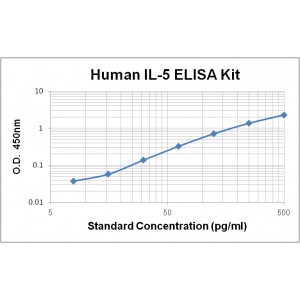
Assay Range | 7.8 - 500 pg/mL |
Sensitivity | 2.0 pg/mL |
Size | 96T |
Storage | Store at 2 - 8ºC. Keep reconstituted standard and detection Ab at -20 ºC |
Assay Principle | Sandwich ELISA |
Sample Volume | 100 µL final volume, dilution factor varies on samples |
Detection Method | Chromogenic |
Kit Components
1. Recombinant Human IL-5 standard: 2 vials
2. One 96-well plate coated with Human IL-5 Ab
3. Sample diluent buffer: 12 mL - 1
4. Detection antibody: 130 µL, dilution 1:100
5. Streptavidin-HRP: 130 µL, dilution 1:100
6. Antibody diluent buffer: 12 mL x1
7. Streptavidin-HRP diluent buffer: 12 mL x1
8. TMB developing agent: 10 mL x1
9. Stop solution: 10 mL x1
10. Washing solution (20x): 25 mL x1
Background
Interleukin 5 (IL-5), also known as B-cell differentiation factor I, Eosinophil differentiation factor, T-cell replacing factor (TRF), is a secreted glycoprotein that belongs to the α-helical group of cytokines including IL-3, IL-5, and GM-CSF. IL-5 is primarily expressed by CD4+ Th2 cells, while activated eosinophils and mast cells can also secreted it with lower amounts. Human IL-5 is 70% identical to mouse IL-5. Human IL-5 receptor consists of a unique ligand-binding subunit (IL-5 Rα) and a common signal-transducing subunit (βc) shared by IL-3 and GM-CSF receptors. Both subunits are members of the cytokine receptor superfamily. IL-5 Rα first binds IL-5 at low affinity and then associates with the βc dimers to form a high-affinity receptor complex containing at least two of each subunit. In addition to the membrane-bound IL-5 Rα subunit, two soluble isoforms of IL-5 Rα have been identified in mouse and human cells. The soluble form of IL-5 Rα is predominant in CD34+ human umbilical cord cells and has been shown to function as an IL-5 antagonist in vitro. Humans IL-5 plays an important role in differentiation, maturation, activation, migration, and survival of eosinophils. Interleukin-5 has been associated with the pathogenesis of several allergic diseases including allergic rhinitis and asthma, in which a dramatic increase of circulating, airway tissue, and induced sputum eosinophils has been observed.


