Competition ELISA Kit Instruction
Kit name and catalog number
Your analyte ELISA Kit, Catalog#: *****
Intended use
The kit is used to detect the level of Your analyte in cell culture, serum, blood plasma and other suitable sample solution.
Assay principle
The coated well immunoenzymatic assay for the quantitative measurement of analyte utilizes a multiclonal anti‐analyte antibody and an analyte‐HRP conjugate. The assay sample and buffer are incubated together with analyte‐HRP conjugate in pre‐coated plate for one hour. After the incubation period, the wells are decanted and washed five times. The wells are then incubated with a substrate for HRP enzyme. The product of the enzyme‐substrate reaction forms a blue colored complex. Finally, a stop solution is added to stop the reaction, which will then turn the solution yellow. The intensity of color is measured spectrophotometrically at 450nm in a microplate reader. The intensity of the color is inversely proportional to the analyte concentration since analyte from samples and analyte ‐HRP conjugate compete for the anti‐ analyte antibody binding site. Since the number of sites is limited, as more sites are occupied by analyanalytete from the sample, fewer sites are left to bind analyte‐HRP conjugate. Standards of known analyte concentrations are run concurrently with the samples being assayed and a standard curve is plotted relating the intensity of the color (Optical Density) to the concentration of analyte. The analyte concentration in each sample is interpolated from this standard curve.
Manufactured and distributed by:
NovaTein Biosciences 310 West Cummings Park Woburn, MA 01801 US
Telephone: (888) 856‐2858
Web: www.novateinbio.com
Materialssupplied
| 1 | Microelisa Stripplate | 96 well | 6 | Chromogenic Substrate A | 6ml X 1vial |
| 2 | Standard | 1.0 ml X 6 vials | 7 | Chromogenic Substrate B | 6ml X 1 vial |
| 3 | 100 X Wash Solution | 10ml X 1vial | 8 | Stop Solution | 6ml X 1 via |
| 4 | Lysis Buffer Solution | 6ml X 1vial | 9 | Specification | 1 |
| 5 | 5 HRP‐Conjugate Reagent | 6ml X 1 vial |
Sample collection and storage
- Serum‐Use a serum separator tube (SST) and allow samples to clot for 30 minutes before a centrifugation for 15minutes at approximately 1000 x g. Remove serum and perform the assay immediately or aliquot and store samples at ‐20 °C or ‐80 °C.
- Plasma‐Collect plasma using EDTA or heparin as an anticoagulant. Centrifuge samples for 15 minutes at 1000 x g at 2‐8 °C within 30minutes of collection. Store samples at ‐20°C or ‐80°C.Avoid repeated freeze‐thaw cycles.
- Cell culture fluid and other biological fluids‐Remove particulates by centrifugation and assay immediately or aliquot and store samples at ‐20°C or ‐80°C. Avoid repeated freeze‐thaw cycles.
- NOTE: The Lysis Buffer Solution is used only when the sample is cell culture fluid & body fluid & tissue homogenate; if the sample is serum or blood plasma, then the Lysis Buffer Solution is a superfluous reagent. Serum, plasma, and cell culture fluid samples to be used within 7 days may be stored at 2‐8 °C, otherwise samples must be stored at ‐20°C(≤2months) or ‐80°C(≤6months) to avoid loss of bioactivity and contamination. Avoid freeze‐thaw cycles. When performing the assay, warm up samples to room temperature slowly. DO NOT USE HEAT‐TREATED SAMPLES.
Materialsrequired but notsupplied
- 1.37 °C incubator
- 2.Standard plate reader capable of measuring absorbance at 450 nm.
- 3.Precision pipettes and disposable pipette tips
- 4.Distilled water
- 5.Multi‐channel pipettes, manifold dispenser or automated microplate washer.
- 6.Absorbent paper
Sample Preparation
- 1.Novateinbio is only responsible for the kit itself, but not for the samples consumed during the assay. The user should calculate the possible amount of the samples used in the whole test. Please reserve sufficient amount ofsamplesin advance.
- 2.Please predict the concentration before assaying. If values for these are not within the range of the standard curve, users must determine the optimal sample dilutions for their particular experiments.
- 3.If the samples are not indicated in the manual, a preliminary experiment to determine the validity ofthe kitis necessary.
- 4 Owing to the possibility of mismatching between antigen from other resource and antibody used in our kits (e.g., antibody targets conformational epitope rather than linear epitope), some native or recombinant proteinsfrom other manufacturers may not be recognized by our products.
- 5.Influenced by the factorsincluding cell viability, cell number and also sampling time,samplesfrom cell culture supernatant may not be detected by the kit
- 6.Fresh samples without long time storage is recommended for the test. Otherwise, protein degradation and denaturalization may occurin those samples and finally lead to wrong results.
Reagent Preparation
- 1.Bring all kit components and samplesto room temperature (18‐25 ℃) before use.
- 2.Dispense 10 μl of Lysis Buffer Solution into 100μl specimens, mix and stand for one hour (The proportion of Lysis Buffer and Specimens should be no less than 1:10). (NOTE: This step is required when the sample is cell culture fluid & body fluid & tissue homogenate; if the sample is serum or blood plasma,then thisstep should be skipped.)
- 3. Wash Solution ‐ Dilute 10 mL of Wash Solution concentrate (100×) with 990 mL of deionized or distilled water to prepare 1000 mL of Wash Solution (1×)
Assay procedures
Prepare all Standards before starting assay procedure (Please read Reagents Preparation). It is recommended that all Standards and Samples be added in duplicate to the Microtiter Plate.
- 1.Secure the desired numbers of coated wellsin the holder then add 100μl of Standards or Samples to the appropriate well of the antibody pre‐coated Microtiter Plate. Generally, you should get the sample value within the assay arrange without dilution. If samples generate values higher than the highest standard, further dilute the samples with 0.1%BSA in PBS, pH7.4 (without Mg2+, Ca2+) and repeat the assay.
- 2.Add 50μl of Conjugate to each well. Mix well. Mixing well in this step is important. Cover and incubate the plate for 1 hour at 37 °C .
- 3. Wash the Microtiter Plate using one of the specified methodsindicated below: Manual Washing: Remove incubation mixture by aspirating contents of the plate into a sink or proper waste container. Fill in each well completely with diluted wash solution, and then aspirate contents of the plate into a sink or proper waste container. Repeat this procedure five times for a total of FIVE washes. After washing, invert plate, and blot dry by hitting the plate onto absorbent paper or paper towels until no moisture appears. Note: Hold the sides of the plate frame firmly when washing the plate to assure that allstripsremain securely in frame. Complete removal of liquid at each step is essential to good performance. Automated Washing: Wash plate FIVE times with diluted wash solution (350‐400 μl/well/wash) using an auto washer. After washing, dry the plate as above. It isrecommended that the washer be setfor a soaking time of 10 seconds and shaking time of 5 seconds between each wash.
- 4.Add 50μl Chromogenic Substrate A and 50μl Chromogenic Substrate B to each well, subsequently. Cover and incubate for 10 minutes at 20‐25 °C . (Avoid sunlight).
- 5.Add 50μl of Stop Solution to each well. Mix well.
- 6.Read the Optical Density (O.D.) at 450 nm using a microtiter plate reader immediately.
Important notes
- 1.The operation should be carried outin strict accordance with the provided instructions.
- 2.To preserve unused strip‐wells, itshould be stored in the sealed bag.
- 3.Always avoid foaming whenmixing orreconstituting protein solutions.
- 4.Pipette reagents and samplesinto the center of eachwell.
- 5.The samples should be transferred into the assay wells within 15 minutes of dilution.
- 6.We recommended that all standard, testing samples are tested in duplicate to minimize the test errors.
- 7.Please predict the concentration before assaying. If values for these are not within the range of the standard curve, users must determine the optimal sample dilutions for their particular experiments.
- 8.Avoid cross‐contamination by changing tips, using separate reservoirs for each reagent, avoid using the suction head without extensive wash.
- 9.Do not mix the reagents from different batches
- 10.Stop Solution should be added in the same order of the Substrate solution.
- 11.Chromogenic Substrate B is light‐sensitive, please avoid prolonged exposure to light.
- 12.The kit should be kept at 2 ‐ 8 °C and cannot be used after expiration date. Standards to be used within 5 days may be stored at 2‐8℃ , otherwise Standards must be stored at ‐20 °C to avoid loss of bioactivity .
Result calculation
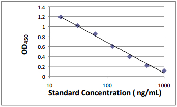
- 1. This standard curve is used to determine the amount of an unknown sample. Construct a standard curve by plotting the average O.D. (450 nm) for each standard on the vertical (Y) axis against the concentration on the horizontal (X) axis, and draw a best fit curve through the points on the graph.
- 2. First, calculate the mean O.D. value for each standard and sample. All O.D. values, are subtracted by the mean value of the blank control before result interpretation. Construct the standard curve using graph paper or statistical software.
- 3. To determine the amount in each sample, first locate the O.D. value on the Y‐axis and extend a horizontal line to the standard curve. At the point of intersection, draw a vertical line to the X‐axis and read the corresponding concentration.
- 4. Any variation in operator, pipetting and washing technique, incubation time or temperature, and kit age can cause variation in result. Each user should obtain their own standard curve.
- 5. Standard curve (reference in general, notforthis kitin particular):
Sensitivity and specificity
- 1. The sensitivity in this assay is 0.1ng/ml (Variable).
- 2. This assay has high sensitivity and excellent specificity for detection of analyte. No significant cross‐reactivity or interference between analyte and analogues was observed.
- 3.Storage: 2‐8 °C
- 4.Validity: six months.