More info
Overview
Long Name | Antibody Type | Antibody Isotype | Host | Species Reactivity | Validated Applications | Purification |
| heparanase | Polyclonal | IgG | Rabbit | Human, Rat | WB | Immunogen affinity purified. |
Immunogen | ||||||
| A synthetic peptide corresponding to a sequence in the middle region of human Heparanase 1 (301-331aa NGRTATKEDFLNPDVLDIFISSVQKVFQVVE), different from the related mouse and rat sequences by eight amino acids. | ||||||
Properties
Form | Lyophilized |
Size | 100 µg/vial |
Contents | Antibody is lyophilized with 5 mg BSA, 0.9 mg NaCl, 0.2 mg Na2HPO4, 0.05 mg NaN3. *carrier free antibody available upon request. |
Concentration | Reconstitute with 0.2 mL sterile dH2O (500 µg/ml final concentration). |
Storage | At -20 °C for 12 months, as supplied. Store reconstituted antibody at 2-8 °C for one month. For long-term storage, aliquot and store at -20 °C. Avoid repeated freezing and thawing. |
Additional Information Regarding the Antigen
Gene | HPSE |
Protein | Heparanase |
Uniprot ID | Q9Y251 |
Function | Endoglycosidase that cleaves heparan sulfate proteoglycans (HSPGs) into heparan sulfate side chains and core proteoglycans. Participates in extracellular matrix (ECM) degradation and remodeling. Selectively cleaves the linkage between a glucuronic acid unit and an N-sulfo glucosamine unit carrying either a 3-O-sulfo or a 6-O-sulfo group. Can also cleave the linkage between a glucuronic acid unit and an N-sulfo glucosamine unit carrying a 2-O-sulfo group, but not linkages between a glucuronic acid unit and a 2-O-sulfated iduronic acid moiety. It is essentially inactive at neutral pH but becomes active under acidic conditions such as during tumor invasion and in inflammatory processes. Facilitates cell migration associated with metastasis, wound healing and inflammation. Enhances shedding of syndecans, and increases endothelial invasion and angiogenesis in myelomas. Acts as procoagulant by increasing the generation of activation factor X in the presence of tissue factor and activation factor VII. Increases cell adhesion to the extacellular matrix (ECM), independent of its enzymatic activity. Induces AKT1/PKB phosphorylation via lipid rafts increasing cell mobility and invasion. Heparin increases this AKT1/PKB activation. Regulates osteogenesis. Enhances angiogenesis through up- regulation of SRC-mediated activation of VEGF. Implicated in hair follicle inner root sheath differentiation and hair homeostasis. |
Tissue Specificity | Highly expressed in placenta and spleen and weakly expressed in lymph node, thymus, peripheral blood leukocytes, bone marrow, endothelial cells, fetal liver and tumor tissues. Also expressed in hair follicles, specifically in both Henle's and Huxley's layers of inner the root sheath (IRS) at anagen phase. |
Sub-cellular localization | Lysosome membrane; Peripheral membrane protein. Secreted. Nucleus. Note: Proheparanase is secreted via vesicles of the Golgi. Interacts with cell membrane heparan sulfate proteoglycans (HSPGs). Endocytosed and accumulates in endosomes. Transferred to lysosomes where it is proteolytically cleaved to produce the active enzyme. Under certain stimuli, transferred to the cell surface. Associates with lipid rafts. Colocalizes with SDC1 in endosomal/lysosomal vesicles. Accumulates in perinuclear lysosomal vesicles. Heparin retains proheparanase in the extracellular medium (By similarity). |
Sequence Similarities | |
Aliases | Endo glucoronidase antibody|Endo-glucoronidase antibody|HEP antibody| Heparanase 50 kDa subunit antibody|Heparanase antibody|Heparanase-1 antibody|Heparanase1 antibody|Hpa 1 antibody|HPA antibody|Hpa1 antibody| HPR 1 antibody|HPR1 antibody|HPSE 1 antibody|HPSE antibody|HPSE_HUMAN antibody|HPSE1 antibody|HSE 1 antibody|HSE1 antibody |
Application Details
| Application | Concentration* | Species | Validated Using** |
| Western blot | 0.1-0.5μg/ml | Human, Rat | AssaySolutio's ECL kit |
AssaySolution recommends Rabbit Chemiluminescent WB Detection Kit (AKIT001B) for Western blot. *Blocking peptide can be purchased at $65. Contact us for more information

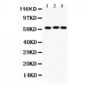
Anti- Heparanase 1 antibody, ASA-B0853, Western blotting
All lanes: Anti Heparanase 1 (ASA-B0853) at 0.5ug/ml
Lane 1: Rat Liver Tissue Lysate at 50ug
Lane 2: Human Placenta Tissue Lysate at 50ug
Lane 3: A549 Whole Cell Lysate at 40ug
Predicted bind size: 61KD
Observed bind size: 61KD
All lanes: Anti Heparanase 1 (ASA-B0853) at 0.5ug/ml
Lane 1: Rat Liver Tissue Lysate at 50ug
Lane 2: Human Placenta Tissue Lysate at 50ug
Lane 3: A549 Whole Cell Lysate at 40ug
Predicted bind size: 61KD
Observed bind size: 61KD


